Sodium »
PDB 6mfm-6myw »
6mrf »
Sodium in PDB 6mrf: Crystal Structure of A Methionine Aminopeptidase Metap From Acinetobacter Baumannii
Enzymatic activity of Crystal Structure of A Methionine Aminopeptidase Metap From Acinetobacter Baumannii
All present enzymatic activity of Crystal Structure of A Methionine Aminopeptidase Metap From Acinetobacter Baumannii:
3.4.11.18;
3.4.11.18;
Protein crystallography data
The structure of Crystal Structure of A Methionine Aminopeptidase Metap From Acinetobacter Baumannii, PDB code: 6mrf
was solved by
Seattle Structural Genomics Center For Infectious Disease (Ssgcid),
with X-Ray Crystallography technique. A brief refinement statistics is given in the table below:
| Resolution Low / High (Å) | 44.73 / 1.65 |
| Space group | P 21 21 21 |
| Cell size a, b, c (Å), α, β, γ (°) | 52.770, 53.690, 84.270, 90.00, 90.00, 90.00 |
| R / Rfree (%) | 16.8 / 21.7 |
Sodium Binding Sites:
The binding sites of Sodium atom in the Crystal Structure of A Methionine Aminopeptidase Metap From Acinetobacter Baumannii
(pdb code 6mrf). This binding sites where shown within
5.0 Angstroms radius around Sodium atom.
In total only one binding site of Sodium was determined in the Crystal Structure of A Methionine Aminopeptidase Metap From Acinetobacter Baumannii, PDB code: 6mrf:
In total only one binding site of Sodium was determined in the Crystal Structure of A Methionine Aminopeptidase Metap From Acinetobacter Baumannii, PDB code: 6mrf:
Sodium binding site 1 out of 1 in 6mrf
Go back to
Sodium binding site 1 out
of 1 in the Crystal Structure of A Methionine Aminopeptidase Metap From Acinetobacter Baumannii
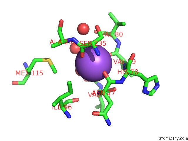
Mono view
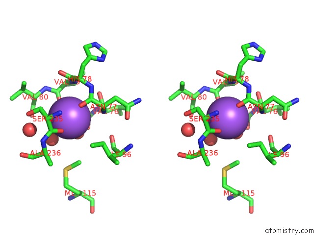
Stereo pair view
Mono view
Stereo pair view
A full contact list of Sodium with other atoms in the Na binding
site number 1 of Crystal Structure of A Methionine Aminopeptidase Metap From Acinetobacter Baumannii within 5.0Å range:
|
Reference:
T.E.Edwards,
S.J.Mayclin,
D.D.Lorimer,
P.S.Horanyi,
Seattle Structural Genomics Center For Infectious Disease.
Crystal Structure of A Methionine Aminopeptidase Metap From Acinetobacter Baumannii To Be Published.
Page generated: Mon Aug 18 06:03:37 2025
Last articles
Zn in 3HBVZn in 3H9Q
Zn in 3HB2
Zn in 3HAY
Zn in 3HAX
Zn in 3H9J
Zn in 3H9G
Zn in 3H8G
Zn in 3H9C
Zn in 3H9B