Sodium »
PDB 6kcd-6lfw »
6kmm »
Sodium in PDB 6kmm: Crystal Structure of Hepes Bound Dye Decolorizing Peroxidase From Bacillus Subtilis
Protein crystallography data
The structure of Crystal Structure of Hepes Bound Dye Decolorizing Peroxidase From Bacillus Subtilis, PDB code: 6kmm
was solved by
P.Dhankhar,
V.Dalal,
J.K.Mahto,
P.Kumar,
with X-Ray Crystallography technique. A brief refinement statistics is given in the table below:
| Resolution Low / High (Å) | 69.91 / 1.93 |
| Space group | P 1 |
| Cell size a, b, c (Å), α, β, γ (°) | 79.837, 101.905, 105.405, 88.00, 76.43, 83.28 |
| R / Rfree (%) | 16.1 / 20.2 |
Other elements in 6kmm:
The structure of Crystal Structure of Hepes Bound Dye Decolorizing Peroxidase From Bacillus Subtilis also contains other interesting chemical elements:
| Iron | (Fe) | 6 atoms |
| Chlorine | (Cl) | 2 atoms |
Sodium Binding Sites:
The binding sites of Sodium atom in the Crystal Structure of Hepes Bound Dye Decolorizing Peroxidase From Bacillus Subtilis
(pdb code 6kmm). This binding sites where shown within
5.0 Angstroms radius around Sodium atom.
In total only one binding site of Sodium was determined in the Crystal Structure of Hepes Bound Dye Decolorizing Peroxidase From Bacillus Subtilis, PDB code: 6kmm:
In total only one binding site of Sodium was determined in the Crystal Structure of Hepes Bound Dye Decolorizing Peroxidase From Bacillus Subtilis, PDB code: 6kmm:
Sodium binding site 1 out of 1 in 6kmm
Go back to
Sodium binding site 1 out
of 1 in the Crystal Structure of Hepes Bound Dye Decolorizing Peroxidase From Bacillus Subtilis
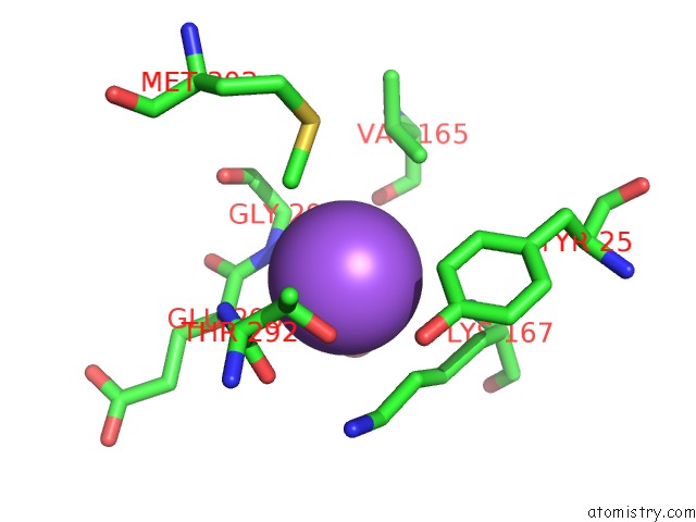
Mono view
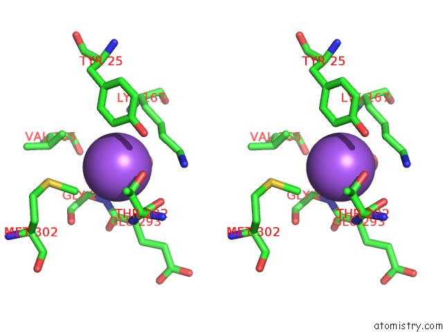
Stereo pair view
Mono view
Stereo pair view
A full contact list of Sodium with other atoms in the Na binding
site number 1 of Crystal Structure of Hepes Bound Dye Decolorizing Peroxidase From Bacillus Subtilis within 5.0Å range:
|
Reference:
P.Dhankhar,
V.Dalal,
J.K.Mahto,
B.R.Gurjar,
S.Tomar,
A.K.Sharma,
P.Kumar.
Characterization of Dye-Decolorizing Peroxidase From Bacillus Subtilis. Arch.Biochem.Biophys. V. 693 08590 2020.
ISSN: ESSN 1096-0384
PubMed: 32971035
DOI: 10.1016/J.ABB.2020.108590
Page generated: Tue Oct 8 11:33:40 2024
ISSN: ESSN 1096-0384
PubMed: 32971035
DOI: 10.1016/J.ABB.2020.108590
Last articles
Zn in 9J0NZn in 9J0O
Zn in 9J0P
Zn in 9FJX
Zn in 9EKB
Zn in 9C0F
Zn in 9CAH
Zn in 9CH0
Zn in 9CH3
Zn in 9CH1