Sodium »
PDB 6f1r-6fmp »
6f7w »
Sodium in PDB 6f7w: Crystal Structure of Dimethylated Rsl - Cucurbit[7]Uril Complex, C2221 Form
Protein crystallography data
The structure of Crystal Structure of Dimethylated Rsl - Cucurbit[7]Uril Complex, C2221 Form, PDB code: 6f7w
was solved by
F.Guagnini,
M.L.Rennie,
P.B.Crowley,
with X-Ray Crystallography technique. A brief refinement statistics is given in the table below:
| Resolution Low / High (Å) | 43.60 / 1.28 |
| Space group | C 2 2 21 |
| Cell size a, b, c (Å), α, β, γ (°) | 50.343, 87.176, 146.595, 90.00, 90.00, 90.00 |
| R / Rfree (%) | 12.4 / 15.1 |
Sodium Binding Sites:
The binding sites of Sodium atom in the Crystal Structure of Dimethylated Rsl - Cucurbit[7]Uril Complex, C2221 Form
(pdb code 6f7w). This binding sites where shown within
5.0 Angstroms radius around Sodium atom.
In total only one binding site of Sodium was determined in the Crystal Structure of Dimethylated Rsl - Cucurbit[7]Uril Complex, C2221 Form, PDB code: 6f7w:
In total only one binding site of Sodium was determined in the Crystal Structure of Dimethylated Rsl - Cucurbit[7]Uril Complex, C2221 Form, PDB code: 6f7w:
Sodium binding site 1 out of 1 in 6f7w
Go back to
Sodium binding site 1 out
of 1 in the Crystal Structure of Dimethylated Rsl - Cucurbit[7]Uril Complex, C2221 Form
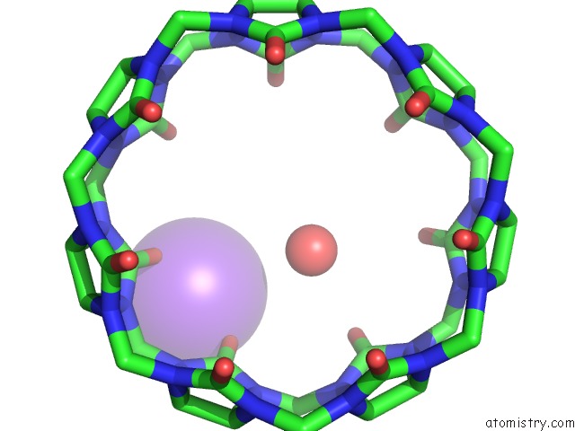
Mono view
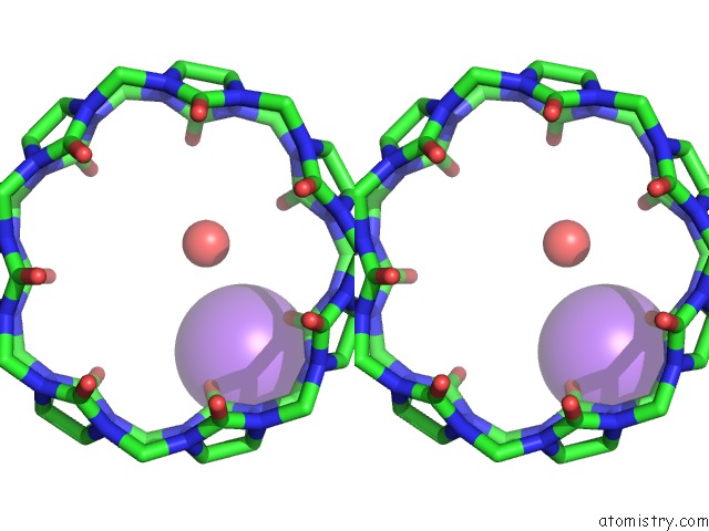
Stereo pair view
Mono view
Stereo pair view
A full contact list of Sodium with other atoms in the Na binding
site number 1 of Crystal Structure of Dimethylated Rsl - Cucurbit[7]Uril Complex, C2221 Form within 5.0Å range:
|
Reference:
F.Guagnini,
P.M.Antonik,
M.L.Rennie,
P.O'byrne,
A.R.Khan,
R.Pinalli,
E.Dalcanale,
P.B.Crowley.
Cucurbit[7]Uril-Dimethyllysine Recognition in A Model Protein. Angew. Chem. Int. Ed. Engl. V. 57 7126 2018.
ISSN: ESSN 1521-3773
PubMed: 29673020
DOI: 10.1002/ANIE.201803232
Page generated: Tue Oct 8 08:42:12 2024
ISSN: ESSN 1521-3773
PubMed: 29673020
DOI: 10.1002/ANIE.201803232
Last articles
Zn in 9J0NZn in 9J0O
Zn in 9J0P
Zn in 9FJX
Zn in 9EKB
Zn in 9C0F
Zn in 9CAH
Zn in 9CH0
Zn in 9CH3
Zn in 9CH1