Sodium »
PDB 6ahh-6awy »
6arc »
Sodium in PDB 6arc: Monoclinic Eutl - Structure Determined From Merged "Group 1" Data
Protein crystallography data
The structure of Monoclinic Eutl - Structure Determined From Merged "Group 1" Data, PDB code: 6arc
was solved by
M.C.Thompson,
D.Cascio,
T.O.Yeates,
with X-Ray Crystallography technique. A brief refinement statistics is given in the table below:
| Resolution Low / High (Å) | 19.80 / 1.90 |
| Space group | C 1 2 1 |
| Cell size a, b, c (Å), α, β, γ (°) | 118.740, 66.140, 80.140, 90.00, 108.53, 90.00 |
| R / Rfree (%) | 13.7 / 18.1 |
Other elements in 6arc:
The structure of Monoclinic Eutl - Structure Determined From Merged "Group 1" Data also contains other interesting chemical elements:
| Chlorine | (Cl) | 1 atom |
Sodium Binding Sites:
The binding sites of Sodium atom in the Monoclinic Eutl - Structure Determined From Merged "Group 1" Data
(pdb code 6arc). This binding sites where shown within
5.0 Angstroms radius around Sodium atom.
In total only one binding site of Sodium was determined in the Monoclinic Eutl - Structure Determined From Merged "Group 1" Data, PDB code: 6arc:
In total only one binding site of Sodium was determined in the Monoclinic Eutl - Structure Determined From Merged "Group 1" Data, PDB code: 6arc:
Sodium binding site 1 out of 1 in 6arc
Go back to
Sodium binding site 1 out
of 1 in the Monoclinic Eutl - Structure Determined From Merged "Group 1" Data
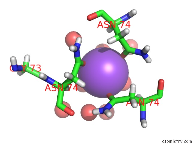
Mono view
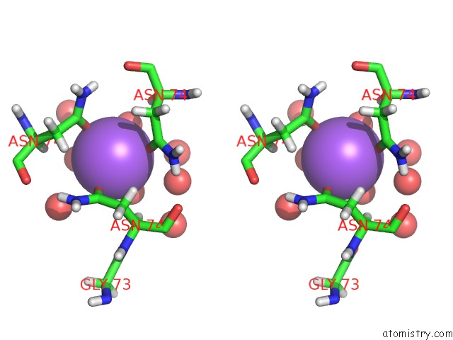
Stereo pair view
Mono view
Stereo pair view
A full contact list of Sodium with other atoms in the Na binding
site number 1 of Monoclinic Eutl - Structure Determined From Merged "Group 1" Data within 5.0Å range:
|
Reference:
M.C.Thompson,
D.Cascio,
T.O.Yeates.
Microfocus Diffraction From Different Regions of A Protein Crystal: Structural Variations and Unit-Cell Polymorphism. Acta Crystallogr D Struct V. 74 411 2018BIOL.
ISSN: ISSN 2059-7983
PubMed: 29717712
DOI: 10.1107/S2059798318003479
Page generated: Tue Oct 8 01:54:37 2024
ISSN: ISSN 2059-7983
PubMed: 29717712
DOI: 10.1107/S2059798318003479
Last articles
Cl in 7T96Cl in 7T94
Cl in 7T8K
Cl in 7T8L
Cl in 7T80
Cl in 7T8J
Cl in 7T88
Cl in 7T4W
Cl in 7T4V
Cl in 7T7K