Sodium »
PDB 5yt7-5zo8 »
5zmi »
Sodium in PDB 5zmi: Crystal Structure of Aprt From Y. Pseudotuberculosis in Complex with Adenine.
Enzymatic activity of Crystal Structure of Aprt From Y. Pseudotuberculosis in Complex with Adenine.
All present enzymatic activity of Crystal Structure of Aprt From Y. Pseudotuberculosis in Complex with Adenine.:
2.4.2.7;
2.4.2.7;
Protein crystallography data
The structure of Crystal Structure of Aprt From Y. Pseudotuberculosis in Complex with Adenine., PDB code: 5zmi
was solved by
G.C.Pavithra,
G.C.Fox,
U.A.Ramagopal,
with X-Ray Crystallography technique. A brief refinement statistics is given in the table below:
| Resolution Low / High (Å) | 48.57 / 2.05 |
| Space group | C 1 2 1 |
| Cell size a, b, c (Å), α, β, γ (°) | 58.958, 78.650, 53.679, 90.00, 115.20, 90.00 |
| R / Rfree (%) | 18.6 / 22.8 |
Sodium Binding Sites:
The binding sites of Sodium atom in the Crystal Structure of Aprt From Y. Pseudotuberculosis in Complex with Adenine.
(pdb code 5zmi). This binding sites where shown within
5.0 Angstroms radius around Sodium atom.
In total only one binding site of Sodium was determined in the Crystal Structure of Aprt From Y. Pseudotuberculosis in Complex with Adenine., PDB code: 5zmi:
In total only one binding site of Sodium was determined in the Crystal Structure of Aprt From Y. Pseudotuberculosis in Complex with Adenine., PDB code: 5zmi:
Sodium binding site 1 out of 1 in 5zmi
Go back to
Sodium binding site 1 out
of 1 in the Crystal Structure of Aprt From Y. Pseudotuberculosis in Complex with Adenine.
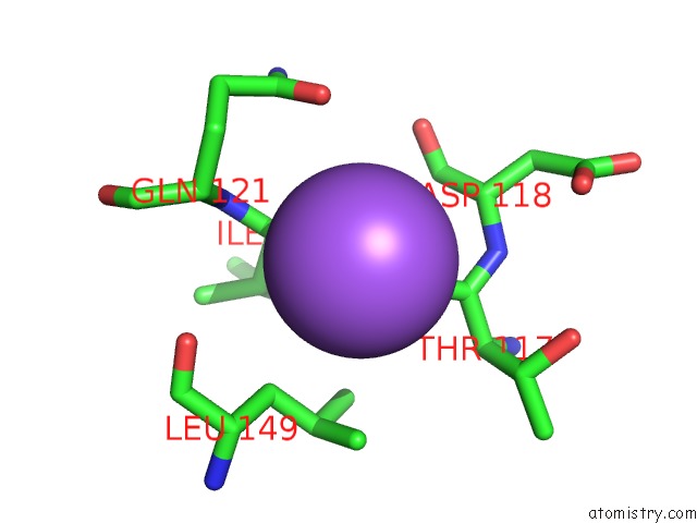
Mono view
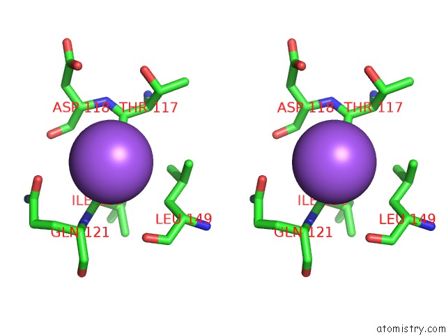
Stereo pair view
Mono view
Stereo pair view
A full contact list of Sodium with other atoms in the Na binding
site number 1 of Crystal Structure of Aprt From Y. Pseudotuberculosis in Complex with Adenine. within 5.0Å range:
|
Reference:
G.C.Pavithra,
U.A.Ramagopal.
Crystal Structure of Adenine Phosphoribosyltransferase From Yersinia Pseudotuberculosis To Be Published.
Page generated: Tue Oct 8 01:31:21 2024
Last articles
Cl in 5FZICl in 5FZH
Cl in 5FZG
Cl in 5FZF
Cl in 5FZE
Cl in 5FZD
Cl in 5FZC
Cl in 5FZB
Cl in 5FZA
Cl in 5FZ9