Sodium »
PDB 5yt7-5zo8 »
5z39 »
Sodium in PDB 5z39: Crystal Structure of C Terminal Region of G-Protein Interacting Protein 1 (GIP1) From Dictyostelium Discoideum Form II
Protein crystallography data
The structure of Crystal Structure of C Terminal Region of G-Protein Interacting Protein 1 (GIP1) From Dictyostelium Discoideum Form II, PDB code: 5z39
was solved by
T.Miyagawa,
H.Koteishi,
Y.Kamimura,
Y.Miyanaga,
K.Takeshita,
A.Nakagawa,
M.Ueda,
with X-Ray Crystallography technique. A brief refinement statistics is given in the table below:
| Resolution Low / High (Å) | 40.09 / 2.74 |
| Space group | P 21 21 21 |
| Cell size a, b, c (Å), α, β, γ (°) | 33.468, 43.618, 101.681, 90.00, 90.00, 90.00 |
| R / Rfree (%) | 22.1 / 27 |
Sodium Binding Sites:
The binding sites of Sodium atom in the Crystal Structure of C Terminal Region of G-Protein Interacting Protein 1 (GIP1) From Dictyostelium Discoideum Form II
(pdb code 5z39). This binding sites where shown within
5.0 Angstroms radius around Sodium atom.
In total only one binding site of Sodium was determined in the Crystal Structure of C Terminal Region of G-Protein Interacting Protein 1 (GIP1) From Dictyostelium Discoideum Form II, PDB code: 5z39:
In total only one binding site of Sodium was determined in the Crystal Structure of C Terminal Region of G-Protein Interacting Protein 1 (GIP1) From Dictyostelium Discoideum Form II, PDB code: 5z39:
Sodium binding site 1 out of 1 in 5z39
Go back to
Sodium binding site 1 out
of 1 in the Crystal Structure of C Terminal Region of G-Protein Interacting Protein 1 (GIP1) From Dictyostelium Discoideum Form II
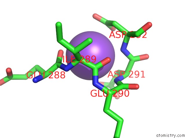
Mono view
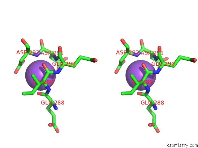
Stereo pair view
Mono view
Stereo pair view
A full contact list of Sodium with other atoms in the Na binding
site number 1 of Crystal Structure of C Terminal Region of G-Protein Interacting Protein 1 (GIP1) From Dictyostelium Discoideum Form II within 5.0Å range:
|
Reference:
T.Miyagawa,
H.Koteishi,
Y.Kamimura,
Y.Miyanaga,
K.Takeshita,
A.Nakagawa,
M.Ueda.
Structural Basis of GIP1 For Cytosolic Sequestration of G Protein in Wide-Range Chemotaxis Nat Commun V. 9 4635 2018.
ISSN: ESSN 2041-1723
PubMed: 30401901
DOI: 10.1038/S41467-018-07035-X
Page generated: Tue Oct 8 01:28:21 2024
ISSN: ESSN 2041-1723
PubMed: 30401901
DOI: 10.1038/S41467-018-07035-X
Last articles
Zn in 9MJ5Zn in 9HNW
Zn in 9G0L
Zn in 9FNE
Zn in 9DZN
Zn in 9E0I
Zn in 9D32
Zn in 9DAK
Zn in 8ZXC
Zn in 8ZUF