Sodium »
PDB 5oqw-5pol »
5osi »
Sodium in PDB 5osi: Structure of Retromer VPS29-VPS35C Subunits Complexed with Ridl Harpin Loop (163-176)
Protein crystallography data
The structure of Structure of Retromer VPS29-VPS35C Subunits Complexed with Ridl Harpin Loop (163-176), PDB code: 5osi
was solved by
M.Romano-Moreno,
A.L.Rojas,
M.Lucas,
M.N.Isupov,
A.Hierro,
with X-Ray Crystallography technique. A brief refinement statistics is given in the table below:
| Resolution Low / High (Å) | 126.49 / 2.52 |
| Space group | P 1 21 1 |
| Cell size a, b, c (Å), α, β, γ (°) | 58.554, 138.993, 126.637, 90.00, 92.78, 90.00 |
| R / Rfree (%) | 20.5 / 26.7 |
Sodium Binding Sites:
The binding sites of Sodium atom in the Structure of Retromer VPS29-VPS35C Subunits Complexed with Ridl Harpin Loop (163-176)
(pdb code 5osi). This binding sites where shown within
5.0 Angstroms radius around Sodium atom.
In total 4 binding sites of Sodium where determined in the Structure of Retromer VPS29-VPS35C Subunits Complexed with Ridl Harpin Loop (163-176), PDB code: 5osi:
Jump to Sodium binding site number: 1; 2; 3; 4;
In total 4 binding sites of Sodium where determined in the Structure of Retromer VPS29-VPS35C Subunits Complexed with Ridl Harpin Loop (163-176), PDB code: 5osi:
Jump to Sodium binding site number: 1; 2; 3; 4;
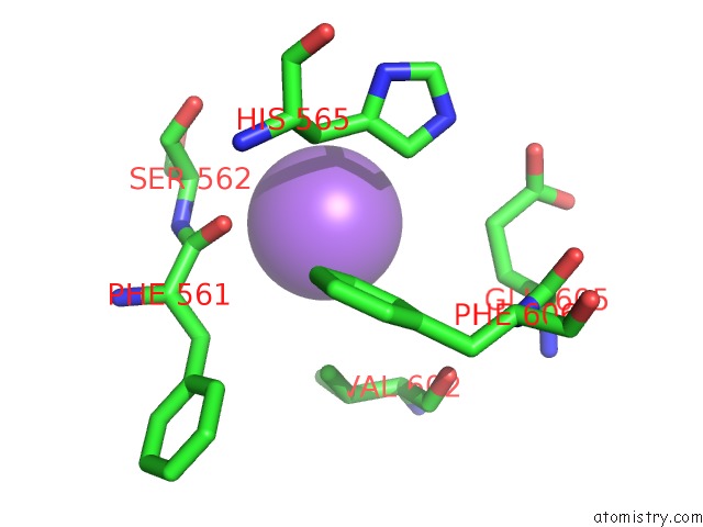
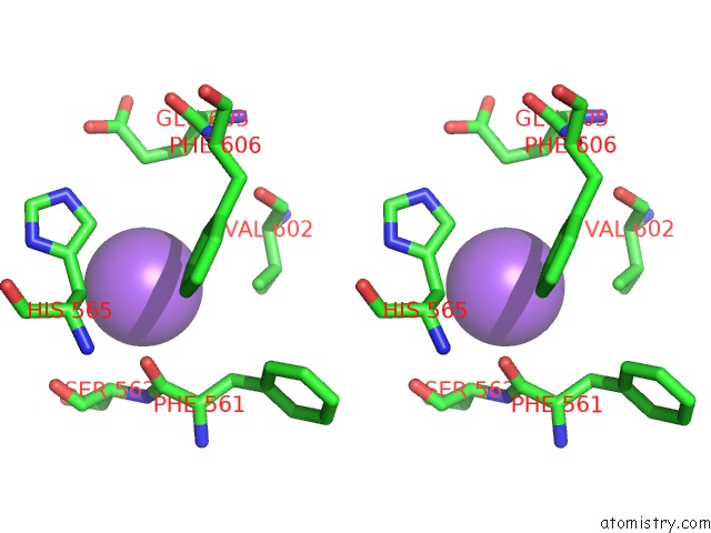
Sodium binding site 1 out of 4 in 5osi
Go back to
Sodium binding site 1 out
of 4 in the Structure of Retromer VPS29-VPS35C Subunits Complexed with Ridl Harpin Loop (163-176)
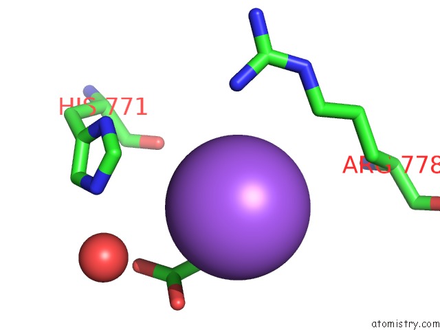
Mono view
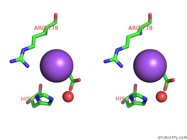
Stereo pair view
Mono view
Stereo pair view
A full contact list of Sodium with other atoms in the Na binding
site number 1 of Structure of Retromer VPS29-VPS35C Subunits Complexed with Ridl Harpin Loop (163-176) within 5.0Å range:
|
Sodium binding site 2 out of 4 in 5osi
Go back to
Sodium binding site 2 out
of 4 in the Structure of Retromer VPS29-VPS35C Subunits Complexed with Ridl Harpin Loop (163-176)
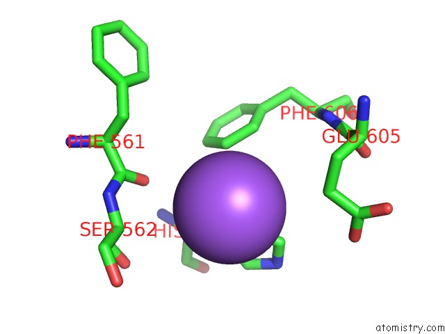
Mono view
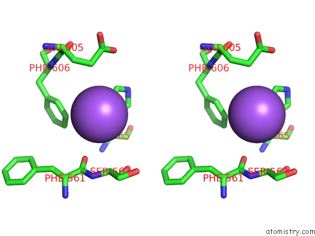
Stereo pair view
Mono view
Stereo pair view
A full contact list of Sodium with other atoms in the Na binding
site number 2 of Structure of Retromer VPS29-VPS35C Subunits Complexed with Ridl Harpin Loop (163-176) within 5.0Å range:
|
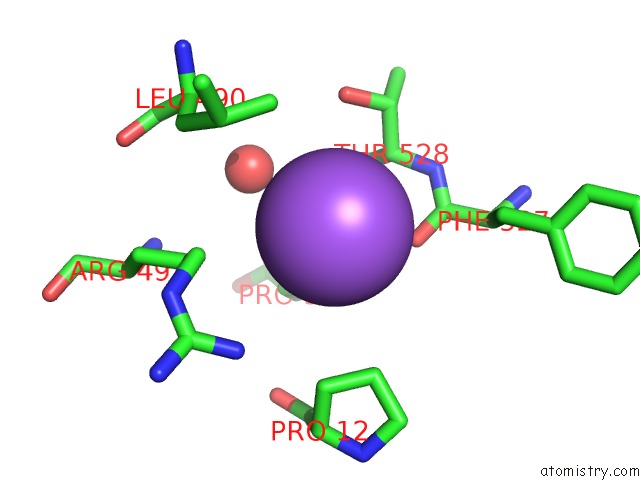
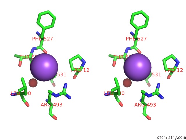
Sodium binding site 3 out of 4 in 5osi
Go back to
Sodium binding site 3 out
of 4 in the Structure of Retromer VPS29-VPS35C Subunits Complexed with Ridl Harpin Loop (163-176)
Mono view
Stereo pair view
Mono view
Stereo pair view
A full contact list of Sodium with other atoms in the Na binding
site number 3 of Structure of Retromer VPS29-VPS35C Subunits Complexed with Ridl Harpin Loop (163-176) within 5.0Å range:
|
Sodium binding site 4 out of 4 in 5osi
Go back to
Sodium binding site 4 out
of 4 in the Structure of Retromer VPS29-VPS35C Subunits Complexed with Ridl Harpin Loop (163-176)
Mono view
Stereo pair view
Mono view
Stereo pair view
A full contact list of Sodium with other atoms in the Na binding
site number 4 of Structure of Retromer VPS29-VPS35C Subunits Complexed with Ridl Harpin Loop (163-176) within 5.0Å range:
|
Reference:
M.Romano-Moreno,
A.L.Rojas,
C.D.Williamson,
D.C.Gershlick,
M.Lucas,
M.N.Isupov,
J.S.Bonifacino,
M.P.Machner,
A.Hierro.
Molecular Mechanism For the Subversion of the Retromer Coat By the Legionella Effector Ridl. Proc. Natl. Acad. Sci. V. 114 11151 2017U.S.A..
ISSN: ESSN 1091-6490
PubMed: 29229824
DOI: 10.1073/PNAS.1715361115
Page generated: Mon Oct 7 23:15:07 2024
ISSN: ESSN 1091-6490
PubMed: 29229824
DOI: 10.1073/PNAS.1715361115
Last articles
Zn in 9MJ5Zn in 9HNW
Zn in 9G0L
Zn in 9FNE
Zn in 9DZN
Zn in 9E0I
Zn in 9D32
Zn in 9DAK
Zn in 8ZXC
Zn in 8ZUF