Sodium »
PDB 5nci-5npm »
5nii »
Sodium in PDB 5nii: Crystal Structure of the Atypical Thioredoxin Reductase Tri From Desulfovibrio Vulgaris Hildenborough
Protein crystallography data
The structure of Crystal Structure of the Atypical Thioredoxin Reductase Tri From Desulfovibrio Vulgaris Hildenborough, PDB code: 5nii
was solved by
O.Valette,
T.T.I.Tran,
C.Cavazza,
E.Caudeville,
G.Brasseur,
A.Dolla,
E.Talla,
L.Pieulle,
with X-Ray Crystallography technique. A brief refinement statistics is given in the table below:
| Resolution Low / High (Å) | 47.48 / 2.00 |
| Space group | I 2 2 2 |
| Cell size a, b, c (Å), α, β, γ (°) | 66.387, 148.161, 152.886, 90.00, 90.00, 90.00 |
| R / Rfree (%) | 19.4 / 22.8 |
Other elements in 5nii:
The structure of Crystal Structure of the Atypical Thioredoxin Reductase Tri From Desulfovibrio Vulgaris Hildenborough also contains other interesting chemical elements:
| Chlorine | (Cl) | 4 atoms |
Sodium Binding Sites:
The binding sites of Sodium atom in the Crystal Structure of the Atypical Thioredoxin Reductase Tri From Desulfovibrio Vulgaris Hildenborough
(pdb code 5nii). This binding sites where shown within
5.0 Angstroms radius around Sodium atom.
In total only one binding site of Sodium was determined in the Crystal Structure of the Atypical Thioredoxin Reductase Tri From Desulfovibrio Vulgaris Hildenborough, PDB code: 5nii:
In total only one binding site of Sodium was determined in the Crystal Structure of the Atypical Thioredoxin Reductase Tri From Desulfovibrio Vulgaris Hildenborough, PDB code: 5nii:
Sodium binding site 1 out of 1 in 5nii
Go back to
Sodium binding site 1 out
of 1 in the Crystal Structure of the Atypical Thioredoxin Reductase Tri From Desulfovibrio Vulgaris Hildenborough

Mono view
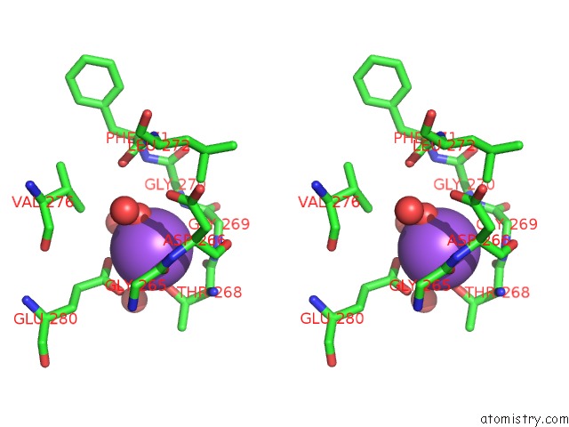
Stereo pair view
Mono view
Stereo pair view
A full contact list of Sodium with other atoms in the Na binding
site number 1 of Crystal Structure of the Atypical Thioredoxin Reductase Tri From Desulfovibrio Vulgaris Hildenborough within 5.0Å range:
|
Reference:
O.Valette,
T.T.T.Tran,
C.Cavazza,
E.Caudeville,
G.Brasseur,
A.Dolla,
E.Talla,
L.Pieulle.
Biochemical Function, Molecular Structure and Evolution of An Atypical Thioredoxin Reductase From Desulfovibrio Vulgaris. Front Microbiol V. 8 1855 2017.
ISSN: ESSN 1664-302X
PubMed: 29033913
DOI: 10.3389/FMICB.2017.01855
Page generated: Mon Oct 7 22:58:44 2024
ISSN: ESSN 1664-302X
PubMed: 29033913
DOI: 10.3389/FMICB.2017.01855
Last articles
Zn in 9J0NZn in 9J0O
Zn in 9J0P
Zn in 9FJX
Zn in 9EKB
Zn in 9C0F
Zn in 9CAH
Zn in 9CH0
Zn in 9CH3
Zn in 9CH1