Sodium »
PDB 5mss-5nbj »
5mxw »
Sodium in PDB 5mxw: Crystal Structure of Yellow Lupin Llpr-10.2B Protein in Complex with Melatonin and Trans-Zeatin.
Protein crystallography data
The structure of Crystal Structure of Yellow Lupin Llpr-10.2B Protein in Complex with Melatonin and Trans-Zeatin., PDB code: 5mxw
was solved by
J.Sliwiak,
M.Sikorski,
M.Jaskolski,
with X-Ray Crystallography technique. A brief refinement statistics is given in the table below:
| Resolution Low / High (Å) | 46.59 / 1.57 |
| Space group | P 65 |
| Cell size a, b, c (Å), α, β, γ (°) | 74.400, 74.400, 67.310, 90.00, 90.00, 120.00 |
| R / Rfree (%) | 18.5 / 20.6 |
Sodium Binding Sites:
The binding sites of Sodium atom in the Crystal Structure of Yellow Lupin Llpr-10.2B Protein in Complex with Melatonin and Trans-Zeatin.
(pdb code 5mxw). This binding sites where shown within
5.0 Angstroms radius around Sodium atom.
In total only one binding site of Sodium was determined in the Crystal Structure of Yellow Lupin Llpr-10.2B Protein in Complex with Melatonin and Trans-Zeatin., PDB code: 5mxw:
In total only one binding site of Sodium was determined in the Crystal Structure of Yellow Lupin Llpr-10.2B Protein in Complex with Melatonin and Trans-Zeatin., PDB code: 5mxw:
Sodium binding site 1 out of 1 in 5mxw
Go back to
Sodium binding site 1 out
of 1 in the Crystal Structure of Yellow Lupin Llpr-10.2B Protein in Complex with Melatonin and Trans-Zeatin.

Mono view
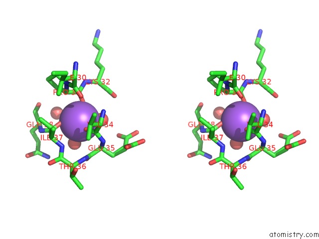
Stereo pair view
Mono view
Stereo pair view
A full contact list of Sodium with other atoms in the Na binding
site number 1 of Crystal Structure of Yellow Lupin Llpr-10.2B Protein in Complex with Melatonin and Trans-Zeatin. within 5.0Å range:
|
Reference:
J.Sliwiak,
M.Sikorski,
M.Jaskolski.
Pr-10 Proteins As Potential Mediators of Melatonin-Cytokinin Cross-Talk in Plants: Crystallographic Studies of Llpr-10.2B Isoform From Yellow Lupine. Febs J. V. 285 1907 2018.
ISSN: ISSN 1742-4658
PubMed: 29630775
DOI: 10.1111/FEBS.14455
Page generated: Mon Oct 7 22:47:46 2024
ISSN: ISSN 1742-4658
PubMed: 29630775
DOI: 10.1111/FEBS.14455
Last articles
Cl in 5RU0Cl in 5RUH
Cl in 5RUO
Cl in 5RUA
Cl in 5RUF
Cl in 5RTX
Cl in 5RUD
Cl in 5RT6
Cl in 5RTH
Cl in 5RTI