Sodium »
PDB 5mft-5mse »
5mk6 »
Sodium in PDB 5mk6: Crystal Structure of the Receptor-Binding Domain of Botulinum Neurotoxin A1 (Crystal Form 1)
Enzymatic activity of Crystal Structure of the Receptor-Binding Domain of Botulinum Neurotoxin A1 (Crystal Form 1)
All present enzymatic activity of Crystal Structure of the Receptor-Binding Domain of Botulinum Neurotoxin A1 (Crystal Form 1):
3.4.24.69;
3.4.24.69;
Protein crystallography data
The structure of Crystal Structure of the Receptor-Binding Domain of Botulinum Neurotoxin A1 (Crystal Form 1), PDB code: 5mk6
was solved by
J.R.Davies,
K.R.Acharya,
with X-Ray Crystallography technique. A brief refinement statistics is given in the table below:
| Resolution Low / High (Å) | 24.05 / 1.45 |
| Space group | P 21 21 21 |
| Cell size a, b, c (Å), α, β, γ (°) | 39.770, 107.320, 107.600, 90.00, 90.00, 90.00 |
| R / Rfree (%) | 17.7 / 22.1 |
Sodium Binding Sites:
The binding sites of Sodium atom in the Crystal Structure of the Receptor-Binding Domain of Botulinum Neurotoxin A1 (Crystal Form 1)
(pdb code 5mk6). This binding sites where shown within
5.0 Angstroms radius around Sodium atom.
In total only one binding site of Sodium was determined in the Crystal Structure of the Receptor-Binding Domain of Botulinum Neurotoxin A1 (Crystal Form 1), PDB code: 5mk6:
In total only one binding site of Sodium was determined in the Crystal Structure of the Receptor-Binding Domain of Botulinum Neurotoxin A1 (Crystal Form 1), PDB code: 5mk6:
Sodium binding site 1 out of 1 in 5mk6
Go back to
Sodium binding site 1 out
of 1 in the Crystal Structure of the Receptor-Binding Domain of Botulinum Neurotoxin A1 (Crystal Form 1)
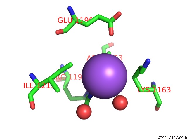
Mono view
Stereo pair view
Mono view
Stereo pair view
A full contact list of Sodium with other atoms in the Na binding
site number 1 of Crystal Structure of the Receptor-Binding Domain of Botulinum Neurotoxin A1 (Crystal Form 1) within 5.0Å range:
|
Reference:
J.R.Davies,
G.S.Hackett,
S.M.Liu,
K.R.Acharya.
High Resolution Crystal Structures of the Receptor-Binding Domain Ofclostridium Botulinumneurotoxin Serotypes A and Fa. Peerj V. 6 E4552 2018.
ISSN: ESSN 2167-8359
PubMed: 29576992
DOI: 10.7717/PEERJ.4552
Page generated: Mon Oct 7 22:42:16 2024
ISSN: ESSN 2167-8359
PubMed: 29576992
DOI: 10.7717/PEERJ.4552
Last articles
Zn in 9J0NZn in 9J0O
Zn in 9J0P
Zn in 9FJX
Zn in 9EKB
Zn in 9C0F
Zn in 9CAH
Zn in 9CH0
Zn in 9CH3
Zn in 9CH1