Sodium »
PDB 5lq7-5m1e »
5m09 »
Sodium in PDB 5m09: Crystal Structure of Mycobacterium Tuberculosis Pkni Kinase Domain, C20A_R136N Double Mutant
Enzymatic activity of Crystal Structure of Mycobacterium Tuberculosis Pkni Kinase Domain, C20A_R136N Double Mutant
All present enzymatic activity of Crystal Structure of Mycobacterium Tuberculosis Pkni Kinase Domain, C20A_R136N Double Mutant:
2.7.11.1;
2.7.11.1;
Protein crystallography data
The structure of Crystal Structure of Mycobacterium Tuberculosis Pkni Kinase Domain, C20A_R136N Double Mutant, PDB code: 5m09
was solved by
M.N.Lisa,
T.Wagner,
M.Alexandre,
N.Barilone,
B.Raynal,
P.M.Alzari,
M.Bellinzoni,
with X-Ray Crystallography technique. A brief refinement statistics is given in the table below:
| Resolution Low / High (Å) | 47.94 / 2.98 |
| Space group | P 43 2 2 |
| Cell size a, b, c (Å), α, β, γ (°) | 110.870, 110.870, 181.760, 90.00, 90.00, 90.00 |
| R / Rfree (%) | 19 / 23.1 |
Sodium Binding Sites:
The binding sites of Sodium atom in the Crystal Structure of Mycobacterium Tuberculosis Pkni Kinase Domain, C20A_R136N Double Mutant
(pdb code 5m09). This binding sites where shown within
5.0 Angstroms radius around Sodium atom.
In total 2 binding sites of Sodium where determined in the Crystal Structure of Mycobacterium Tuberculosis Pkni Kinase Domain, C20A_R136N Double Mutant, PDB code: 5m09:
Jump to Sodium binding site number: 1; 2;
In total 2 binding sites of Sodium where determined in the Crystal Structure of Mycobacterium Tuberculosis Pkni Kinase Domain, C20A_R136N Double Mutant, PDB code: 5m09:
Jump to Sodium binding site number: 1; 2;
Sodium binding site 1 out of 2 in 5m09
Go back to
Sodium binding site 1 out
of 2 in the Crystal Structure of Mycobacterium Tuberculosis Pkni Kinase Domain, C20A_R136N Double Mutant
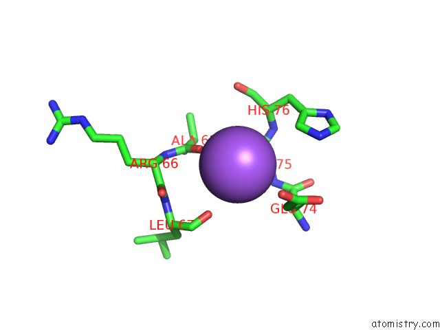
Mono view
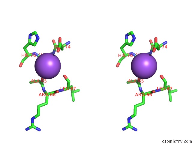
Stereo pair view
Mono view
Stereo pair view
A full contact list of Sodium with other atoms in the Na binding
site number 1 of Crystal Structure of Mycobacterium Tuberculosis Pkni Kinase Domain, C20A_R136N Double Mutant within 5.0Å range:
|
Sodium binding site 2 out of 2 in 5m09
Go back to
Sodium binding site 2 out
of 2 in the Crystal Structure of Mycobacterium Tuberculosis Pkni Kinase Domain, C20A_R136N Double Mutant
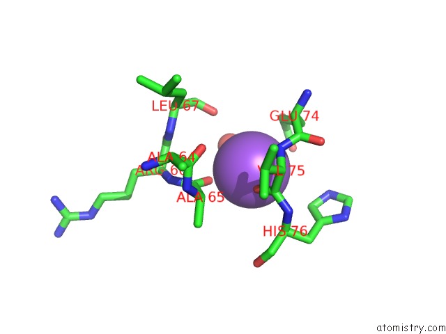
Mono view
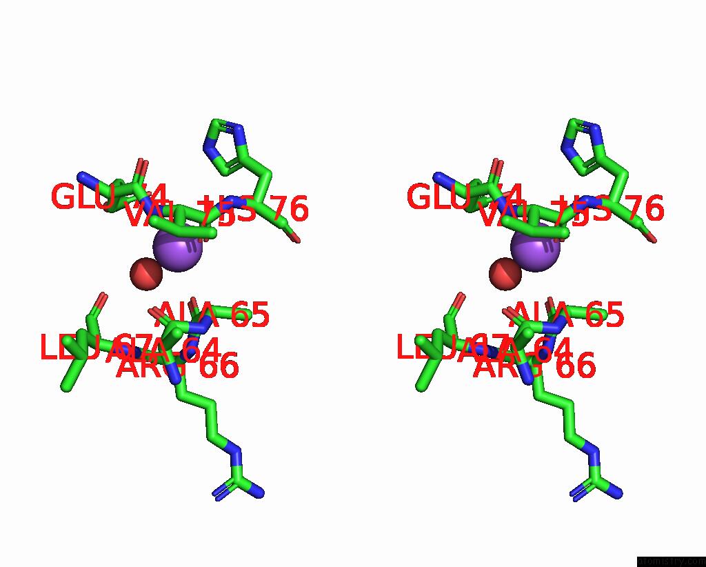
Stereo pair view
Mono view
Stereo pair view
A full contact list of Sodium with other atoms in the Na binding
site number 2 of Crystal Structure of Mycobacterium Tuberculosis Pkni Kinase Domain, C20A_R136N Double Mutant within 5.0Å range:
|
Reference:
M.N.Lisa,
T.Wagner,
M.Alexandre,
N.Barilone,
B.Raynal,
P.M.Alzari,
M.Bellinzoni.
The Crystal Structure of Pkni From Mycobacterium Tuberculosis Shows An Inactive, Pseudokinase-Like Conformation. Febs J. V. 284 602 2017.
ISSN: ISSN 1742-4658
PubMed: 28054744
DOI: 10.1111/FEBS.14003
Page generated: Mon Oct 7 22:27:15 2024
ISSN: ISSN 1742-4658
PubMed: 28054744
DOI: 10.1111/FEBS.14003
Last articles
Zn in 9J0NZn in 9J0O
Zn in 9J0P
Zn in 9FJX
Zn in 9EKB
Zn in 9C0F
Zn in 9CAH
Zn in 9CH0
Zn in 9CH3
Zn in 9CH1