Sodium in PDB 5lx8: Crystal Structure of BT1762
Protein crystallography data
The structure of Crystal Structure of BT1762, PDB code: 5lx8
was solved by
A.Basle,
with X-Ray Crystallography technique. A brief refinement statistics is given in the table below:
Resolution Low / High (Å)
|
45.70 /
1.76
|
Space group
|
C 2 2 21
|
Cell size a, b, c (Å), α, β, γ (°)
|
107.633,
129.380,
86.610,
90.00,
90.00,
90.00
|
R / Rfree (%)
|
15.6 /
18.9
|
Sodium Binding Sites:
The binding sites of Sodium atom in the Crystal Structure of BT1762
(pdb code 5lx8). This binding sites where shown within
5.0 Angstroms radius around Sodium atom.
In total 3 binding sites of Sodium where determined in the
Crystal Structure of BT1762, PDB code: 5lx8:
Jump to Sodium binding site number:
1;
2;
3;
Sodium binding site 1 out
of 3 in 5lx8
Go back to
Sodium Binding Sites List in 5lx8
Sodium binding site 1 out
of 3 in the Crystal Structure of BT1762
Mono view
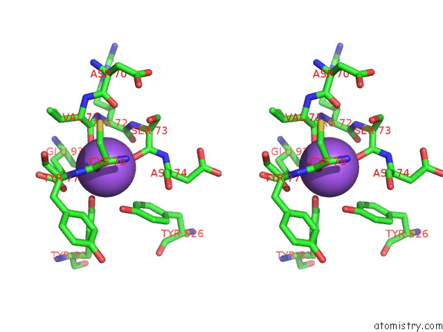
Stereo pair view
|
A full contact list of Sodium with other atoms in the Na binding
site number 1 of Crystal Structure of BT1762 within 5.0Å range:
probe
|
atom
|
residue
|
distance (Å)
|
B
|
Occ
|
A:Na606
b:13.3
occ:1.00
|
O
|
A:CYS76
|
2.3
|
13.2
|
1.0
|
O
|
A:SER73
|
2.3
|
11.5
|
1.0
|
O
|
A:VAL71
|
2.3
|
14.0
|
1.0
|
O
|
A:HOH913
|
2.5
|
13.2
|
1.0
|
OH
|
A:TYR537
|
2.6
|
15.0
|
1.0
|
C
|
A:VAL71
|
3.4
|
12.6
|
1.0
|
C
|
A:CYS76
|
3.5
|
12.9
|
1.0
|
C
|
A:SER73
|
3.5
|
11.3
|
1.0
|
OH
|
A:TYR526
|
3.6
|
13.5
|
1.0
|
OE2
|
A:GLU93
|
3.6
|
15.5
|
1.0
|
CE2
|
A:TYR526
|
3.8
|
11.9
|
1.0
|
CG
|
A:GLU93
|
3.8
|
14.0
|
1.0
|
CZ
|
A:TYR537
|
3.8
|
15.6
|
1.0
|
C
|
A:ARG72
|
3.9
|
12.3
|
1.0
|
N
|
A:SER73
|
4.0
|
11.5
|
1.0
|
CD
|
A:GLU93
|
4.0
|
15.3
|
1.0
|
CZ
|
A:TYR526
|
4.1
|
12.9
|
1.0
|
O
|
A:ARG72
|
4.2
|
13.2
|
1.0
|
CA
|
A:VAL71
|
4.3
|
13.8
|
1.0
|
N
|
A:CYS76
|
4.3
|
12.4
|
1.0
|
N
|
A:ARG72
|
4.3
|
12.3
|
1.0
|
CE1
|
A:TYR537
|
4.3
|
16.5
|
1.0
|
CA
|
A:ARG72
|
4.3
|
12.5
|
1.0
|
CA
|
A:SER73
|
4.4
|
10.8
|
1.0
|
CA
|
A:CYS76
|
4.4
|
14.0
|
1.0
|
N
|
A:TYR77
|
4.5
|
12.2
|
1.0
|
N
|
A:ASP74
|
4.6
|
12.1
|
1.0
|
CA
|
A:TYR77
|
4.6
|
12.5
|
1.0
|
O
|
A:GLU93
|
4.7
|
12.9
|
1.0
|
CA
|
A:ASP74
|
4.8
|
12.0
|
1.0
|
O
|
A:ASP70
|
4.8
|
13.6
|
1.0
|
CD2
|
A:TYR526
|
4.8
|
12.8
|
1.0
|
CD1
|
A:TYR77
|
4.9
|
12.1
|
1.0
|
CB
|
A:GLU93
|
4.9
|
14.0
|
1.0
|
CE2
|
A:TYR537
|
5.0
|
16.4
|
1.0
|
CB
|
A:VAL71
|
5.0
|
14.7
|
1.0
|
|
Sodium binding site 2 out
of 3 in 5lx8
Go back to
Sodium Binding Sites List in 5lx8
Sodium binding site 2 out
of 3 in the Crystal Structure of BT1762
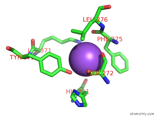
Mono view
Stereo pair view
|
A full contact list of Sodium with other atoms in the Na binding
site number 2 of Crystal Structure of BT1762 within 5.0Å range:
probe
|
atom
|
residue
|
distance (Å)
|
B
|
Occ
|
A:Na607
b:28.1
occ:1.00
|
O
|
A:HOH853
|
2.5
|
14.9
|
1.0
|
OH
|
A:TYR372
|
2.7
|
13.7
|
1.0
|
O
|
A:SER272
|
3.0
|
10.3
|
1.0
|
CB
|
A:PHE275
|
3.5
|
10.8
|
1.0
|
CB
|
A:SER272
|
3.6
|
12.7
|
1.0
|
CE2
|
A:TYR372
|
3.6
|
14.4
|
1.0
|
CZ
|
A:TYR372
|
3.6
|
13.7
|
1.0
|
CA
|
A:SER272
|
3.6
|
11.5
|
1.0
|
C
|
A:SER272
|
3.7
|
10.9
|
1.0
|
NZ
|
A:LYS371
|
3.7
|
13.3
|
1.0
|
O
|
A:PHE275
|
4.0
|
10.9
|
1.0
|
CD2
|
A:PHE275
|
4.0
|
11.2
|
1.0
|
CG
|
A:LEU276
|
4.0
|
13.2
|
1.0
|
CE
|
A:LYS371
|
4.0
|
13.8
|
1.0
|
C
|
A:PHE275
|
4.1
|
12.0
|
1.0
|
CG
|
A:PHE275
|
4.2
|
10.6
|
1.0
|
CA
|
A:PHE275
|
4.3
|
11.1
|
1.0
|
CD2
|
A:LEU276
|
4.3
|
14.1
|
1.0
|
O
|
A:HIS361
|
4.4
|
10.9
|
1.0
|
CD1
|
A:LEU276
|
4.4
|
14.2
|
1.0
|
CB
|
A:HIS361
|
4.5
|
10.8
|
1.0
|
ND1
|
A:HIS361
|
4.5
|
10.4
|
1.0
|
N
|
A:PHE275
|
4.6
|
10.3
|
1.0
|
OG
|
A:SER272
|
4.7
|
14.7
|
1.0
|
N
|
A:LEU276
|
4.8
|
12.0
|
1.0
|
CA
|
A:HIS361
|
4.9
|
10.8
|
1.0
|
CG
|
A:HIS361
|
4.9
|
10.3
|
1.0
|
CD2
|
A:TYR372
|
4.9
|
14.4
|
1.0
|
CE1
|
A:TYR372
|
4.9
|
13.7
|
1.0
|
N
|
A:SER272
|
5.0
|
11.1
|
1.0
|
|
Sodium binding site 3 out
of 3 in 5lx8
Go back to
Sodium Binding Sites List in 5lx8
Sodium binding site 3 out
of 3 in the Crystal Structure of BT1762
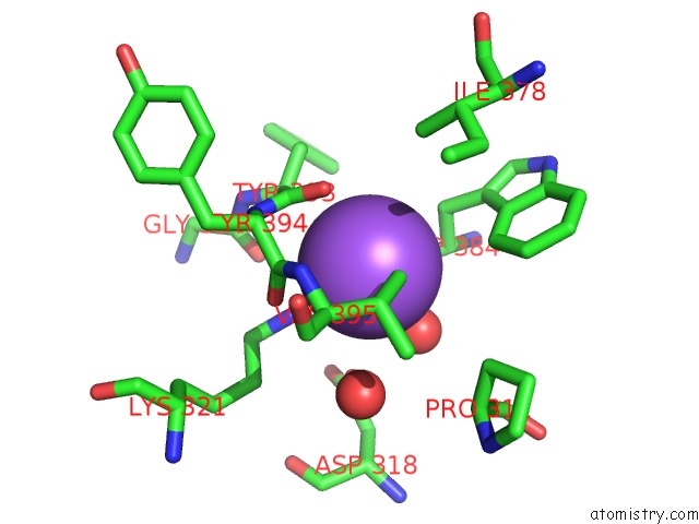
Mono view
Stereo pair view
|
A full contact list of Sodium with other atoms in the Na binding
site number 3 of Crystal Structure of BT1762 within 5.0Å range:
probe
|
atom
|
residue
|
distance (Å)
|
B
|
Occ
|
A:Na608
b:37.0
occ:1.00
|
O
|
A:TRP384
|
2.5
|
19.5
|
1.0
|
O
|
A:HOH755
|
2.6
|
22.8
|
1.0
|
O
|
A:TYR393
|
2.9
|
15.8
|
1.0
|
NZ
|
A:LYS321
|
3.0
|
15.9
|
1.0
|
CG2
|
A:VAL395
|
3.3
|
18.3
|
1.0
|
C
|
A:TYR393
|
3.4
|
14.8
|
1.0
|
C
|
A:TRP384
|
3.4
|
19.7
|
1.0
|
O
|
A:TYR394
|
3.5
|
14.3
|
1.0
|
CB
|
A:TRP384
|
3.6
|
19.2
|
1.0
|
C
|
A:TYR394
|
3.8
|
13.7
|
1.0
|
CA
|
A:TRP384
|
3.8
|
20.1
|
1.0
|
CA
|
A:TYR393
|
3.8
|
15.8
|
1.0
|
OD1
|
A:ASP318
|
3.8
|
17.1
|
1.0
|
N
|
A:TYR394
|
4.0
|
13.7
|
1.0
|
CE3
|
A:TRP384
|
4.1
|
19.0
|
1.0
|
O
|
A:HOH925
|
4.1
|
20.7
|
1.0
|
N
|
A:VAL395
|
4.3
|
13.4
|
1.0
|
CA
|
A:TYR394
|
4.4
|
14.0
|
1.0
|
CB
|
A:VAL395
|
4.5
|
15.7
|
1.0
|
CE
|
A:LYS321
|
4.5
|
16.2
|
1.0
|
CG
|
A:TRP384
|
4.6
|
19.2
|
1.0
|
CA
|
A:VAL395
|
4.6
|
13.6
|
1.0
|
N
|
A:SER385
|
4.6
|
20.6
|
1.0
|
CD2
|
A:TRP384
|
4.6
|
19.8
|
1.0
|
O
|
A:GLY392
|
4.7
|
16.3
|
1.0
|
CB
|
A:TYR393
|
4.7
|
17.4
|
1.0
|
CG2
|
A:ILE378
|
4.8
|
18.4
|
1.0
|
CG
|
A:ASP318
|
4.8
|
17.6
|
1.0
|
N
|
A:TYR393
|
4.9
|
16.1
|
1.0
|
CB
|
A:PRO311
|
5.0
|
19.9
|
1.0
|
|
Reference:
A.J.Glenwright,
K.R.Pothula,
S.P.Bhamidimarri,
D.S.Chorev,
A.Basle,
S.J.Firbank,
H.Zheng,
C.V.Robinson,
M.Winterhalter,
U.Kleinekathofer,
D.N.Bolam,
B.Van Den Berg.
Structural Basis For Nutrient Acquisition By Dominant Members of the Human Gut Microbiota. Nature V. 541 407 2017.
ISSN: ESSN 1476-4687
PubMed: 28077872
DOI: 10.1038/NATURE20828
Page generated: Mon Oct 7 22:26:06 2024
|