Sodium »
PDB 5lq7-5m1e »
5lwq »
Sodium in PDB 5lwq: Ceue (H227L Variant) A Periplasmic Protein From Campylobacter Jejuni
Protein crystallography data
The structure of Ceue (H227L Variant) A Periplasmic Protein From Campylobacter Jejuni, PDB code: 5lwq
was solved by
E.J.Wilde,
E.Blagova,
A.Hughes,
D.J.Raines,
O.V.Moroz,
J.P.Turkenburg,
A.-K.Duhme-Klair,
K.S.Wilson,
with X-Ray Crystallography technique. A brief refinement statistics is given in the table below:
| Resolution Low / High (Å) | 65.52 / 1.52 |
| Space group | P 1 |
| Cell size a, b, c (Å), α, β, γ (°) | 56.921, 62.560, 67.789, 82.21, 76.99, 76.18 |
| R / Rfree (%) | 18.5 / 22.1 |
Other elements in 5lwq:
The structure of Ceue (H227L Variant) A Periplasmic Protein From Campylobacter Jejuni also contains other interesting chemical elements:
| Bromine | (Br) | 5 atoms |
Sodium Binding Sites:
The binding sites of Sodium atom in the Ceue (H227L Variant) A Periplasmic Protein From Campylobacter Jejuni
(pdb code 5lwq). This binding sites where shown within
5.0 Angstroms radius around Sodium atom.
In total only one binding site of Sodium was determined in the Ceue (H227L Variant) A Periplasmic Protein From Campylobacter Jejuni, PDB code: 5lwq:
In total only one binding site of Sodium was determined in the Ceue (H227L Variant) A Periplasmic Protein From Campylobacter Jejuni, PDB code: 5lwq:
Sodium binding site 1 out of 1 in 5lwq
Go back to
Sodium binding site 1 out
of 1 in the Ceue (H227L Variant) A Periplasmic Protein From Campylobacter Jejuni
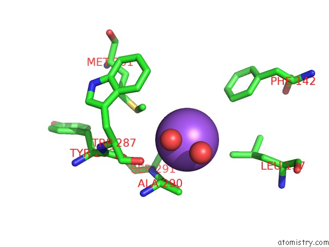
Mono view
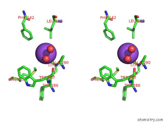
Stereo pair view
Mono view
Stereo pair view
A full contact list of Sodium with other atoms in the Na binding
site number 1 of Ceue (H227L Variant) A Periplasmic Protein From Campylobacter Jejuni within 5.0Å range:
|
Reference:
E.J.Wilde,
A.Hughes,
E.V.Blagova,
O.V.Moroz,
R.P.Thomas,
J.P.Turkenburg,
D.J.Raines,
A.K.Duhme-Klair,
K.S.Wilson.
Interactions of the Periplasmic Binding Protein Ceue with Fe(III) N-Licam(4-) Siderophore Analogues of Varied Linker Length. Sci Rep V. 7 45941 2017.
ISSN: ESSN 2045-2322
PubMed: 28383577
DOI: 10.1038/SREP45941
Page generated: Mon Oct 7 22:26:03 2024
ISSN: ESSN 2045-2322
PubMed: 28383577
DOI: 10.1038/SREP45941
Last articles
Zn in 9MJ5Zn in 9HNW
Zn in 9G0L
Zn in 9FNE
Zn in 9DZN
Zn in 9E0I
Zn in 9D32
Zn in 9DAK
Zn in 8ZXC
Zn in 8ZUF