Sodium »
PDB 5g3g-5gwa »
5ghm »
Sodium in PDB 5ghm: Crystal Structure of Human MTH1(G2K/D120N Mutant) in Complex with 8- Oxo-Dgtp at pH 7.0
Enzymatic activity of Crystal Structure of Human MTH1(G2K/D120N Mutant) in Complex with 8- Oxo-Dgtp at pH 7.0
All present enzymatic activity of Crystal Structure of Human MTH1(G2K/D120N Mutant) in Complex with 8- Oxo-Dgtp at pH 7.0:
3.6.1.55; 3.6.1.56;
3.6.1.55; 3.6.1.56;
Protein crystallography data
The structure of Crystal Structure of Human MTH1(G2K/D120N Mutant) in Complex with 8- Oxo-Dgtp at pH 7.0, PDB code: 5ghm
was solved by
T.Nakamura,
S.Waz,
K.Hirata,
Y.Nakabeppu,
Y.Yamagata,
with X-Ray Crystallography technique. A brief refinement statistics is given in the table below:
| Resolution Low / High (Å) | 37.80 / 1.50 |
| Space group | P 21 21 21 |
| Cell size a, b, c (Å), α, β, γ (°) | 45.790, 47.778, 123.610, 90.00, 90.00, 90.00 |
| R / Rfree (%) | 17.3 / 19.6 |
Sodium Binding Sites:
The binding sites of Sodium atom in the Crystal Structure of Human MTH1(G2K/D120N Mutant) in Complex with 8- Oxo-Dgtp at pH 7.0
(pdb code 5ghm). This binding sites where shown within
5.0 Angstroms radius around Sodium atom.
In total only one binding site of Sodium was determined in the Crystal Structure of Human MTH1(G2K/D120N Mutant) in Complex with 8- Oxo-Dgtp at pH 7.0, PDB code: 5ghm:
In total only one binding site of Sodium was determined in the Crystal Structure of Human MTH1(G2K/D120N Mutant) in Complex with 8- Oxo-Dgtp at pH 7.0, PDB code: 5ghm:
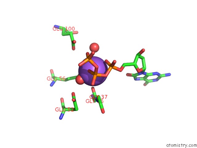
Sodium binding site 1 out of 1 in 5ghm
Go back to
Sodium binding site 1 out
of 1 in the Crystal Structure of Human MTH1(G2K/D120N Mutant) in Complex with 8- Oxo-Dgtp at pH 7.0
Mono view

Stereo pair view
Mono view
Stereo pair view
A full contact list of Sodium with other atoms in the Na binding
site number 1 of Crystal Structure of Human MTH1(G2K/D120N Mutant) in Complex with 8- Oxo-Dgtp at pH 7.0 within 5.0Å range:
|
Reference:
S.Waz,
T.Nakamura,
K.Hirata,
Y.Koga-Ogawa,
M.Chirifu,
T.Arimori,
T.Tamada,
S.Ikemizu,
Y.Nakabeppu,
Y.Yamagata.
Structural and Kinetic Studies of the Human Nudix Hydrolase MTH1 Reveal the Mechanism For Its Broad Substrate Specificity J. Biol. Chem. V. 292 2785 2017.
ISSN: ESSN 1083-351X
PubMed: 28035004
DOI: 10.1074/JBC.M116.749713
Page generated: Mon Oct 7 21:10:18 2024
ISSN: ESSN 1083-351X
PubMed: 28035004
DOI: 10.1074/JBC.M116.749713
Last articles
Fe in 2YXOFe in 2YRS
Fe in 2YXC
Fe in 2YNM
Fe in 2YVJ
Fe in 2YP1
Fe in 2YU2
Fe in 2YU1
Fe in 2YQB
Fe in 2YOO