Sodium »
PDB 5dyn-5emg »
5ek8 »
Sodium in PDB 5ek8: Crystal Structure of A 9R-Lipoxygenase From Cyanothece PCC8801 at 2.7 Angstroms
Protein crystallography data
The structure of Crystal Structure of A 9R-Lipoxygenase From Cyanothece PCC8801 at 2.7 Angstroms, PDB code: 5ek8
was solved by
I.Feussner,
R.Ficner,
P.Neumann,
J.Newie,
A.Andreou,
O.Einsle,
with X-Ray Crystallography technique. A brief refinement statistics is given in the table below:
| Resolution Low / High (Å) | 19.76 / 2.70 |
| Space group | P 64 2 2 |
| Cell size a, b, c (Å), α, β, γ (°) | 121.166, 121.166, 234.673, 90.00, 90.00, 120.00 |
| R / Rfree (%) | 19.9 / 24.6 |
Other elements in 5ek8:
The structure of Crystal Structure of A 9R-Lipoxygenase From Cyanothece PCC8801 at 2.7 Angstroms also contains other interesting chemical elements:
| Iron | (Fe) | 1 atom |
Sodium Binding Sites:
The binding sites of Sodium atom in the Crystal Structure of A 9R-Lipoxygenase From Cyanothece PCC8801 at 2.7 Angstroms
(pdb code 5ek8). This binding sites where shown within
5.0 Angstroms radius around Sodium atom.
In total only one binding site of Sodium was determined in the Crystal Structure of A 9R-Lipoxygenase From Cyanothece PCC8801 at 2.7 Angstroms, PDB code: 5ek8:
In total only one binding site of Sodium was determined in the Crystal Structure of A 9R-Lipoxygenase From Cyanothece PCC8801 at 2.7 Angstroms, PDB code: 5ek8:
Sodium binding site 1 out of 1 in 5ek8
Go back to
Sodium binding site 1 out
of 1 in the Crystal Structure of A 9R-Lipoxygenase From Cyanothece PCC8801 at 2.7 Angstroms
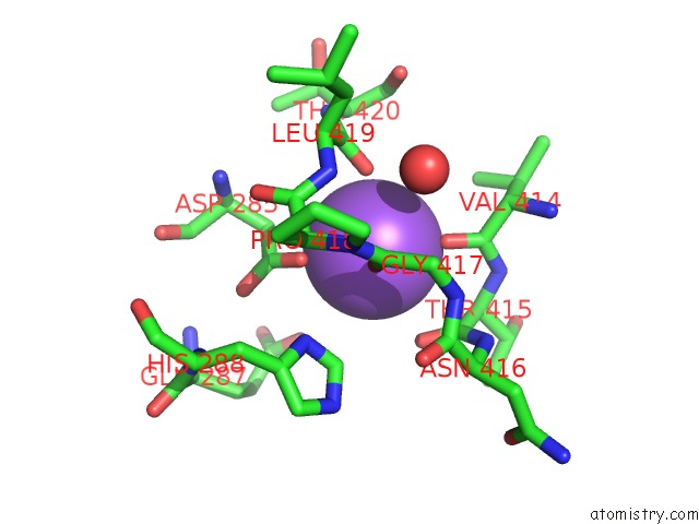
Mono view
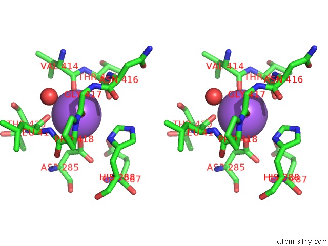
Stereo pair view
Mono view
Stereo pair view
A full contact list of Sodium with other atoms in the Na binding
site number 1 of Crystal Structure of A 9R-Lipoxygenase From Cyanothece PCC8801 at 2.7 Angstroms within 5.0Å range:
|
Reference:
J.Newie,
A.Andreou,
P.Neumann,
O.Einsle,
I.Feussner,
R.Ficner.
Crystal Structure of A Lipoxygenase From Cyanothece Sp. May Reveal Novel Features For Substrate Acquisition. J.Lipid Res. V. 57 276 2016.
ISSN: ISSN 0022-2275
PubMed: 26667668
DOI: 10.1194/JLR.M064980
Page generated: Mon Oct 7 20:48:16 2024
ISSN: ISSN 0022-2275
PubMed: 26667668
DOI: 10.1194/JLR.M064980
Last articles
Zn in 9J0NZn in 9J0O
Zn in 9J0P
Zn in 9FJX
Zn in 9EKB
Zn in 9C0F
Zn in 9CAH
Zn in 9CH0
Zn in 9CH3
Zn in 9CH1