Sodium in PDB 4xnd: Crystal Structure of E. Coli Aminopeptidase N in Complex with L-Beta Homotryptophan
Enzymatic activity of Crystal Structure of E. Coli Aminopeptidase N in Complex with L-Beta Homotryptophan
All present enzymatic activity of Crystal Structure of E. Coli Aminopeptidase N in Complex with L-Beta Homotryptophan:
3.4.11.2;
Protein crystallography data
The structure of Crystal Structure of E. Coli Aminopeptidase N in Complex with L-Beta Homotryptophan, PDB code: 4xnd
was solved by
A.Addlagatta,
R.Gumpena,
with X-Ray Crystallography technique. A brief refinement statistics is given in the table below:
Resolution Low / High (Å)
|
20.00 /
1.93
|
Space group
|
P 31 2 1
|
Cell size a, b, c (Å), α, β, γ (°)
|
121.120,
121.120,
170.781,
90.00,
90.00,
120.00
|
R / Rfree (%)
|
13.3 /
16.6
|
Other elements in 4xnd:
The structure of Crystal Structure of E. Coli Aminopeptidase N in Complex with L-Beta Homotryptophan also contains other interesting chemical elements:
Sodium Binding Sites:
The binding sites of Sodium atom in the Crystal Structure of E. Coli Aminopeptidase N in Complex with L-Beta Homotryptophan
(pdb code 4xnd). This binding sites where shown within
5.0 Angstroms radius around Sodium atom.
In total 8 binding sites of Sodium where determined in the
Crystal Structure of E. Coli Aminopeptidase N in Complex with L-Beta Homotryptophan, PDB code: 4xnd:
Jump to Sodium binding site number:
1;
2;
3;
4;
5;
6;
7;
8;
Sodium binding site 1 out
of 8 in 4xnd
Go back to
Sodium Binding Sites List in 4xnd
Sodium binding site 1 out
of 8 in the Crystal Structure of E. Coli Aminopeptidase N in Complex with L-Beta Homotryptophan
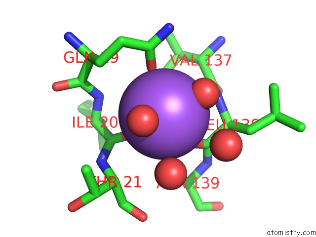
Mono view
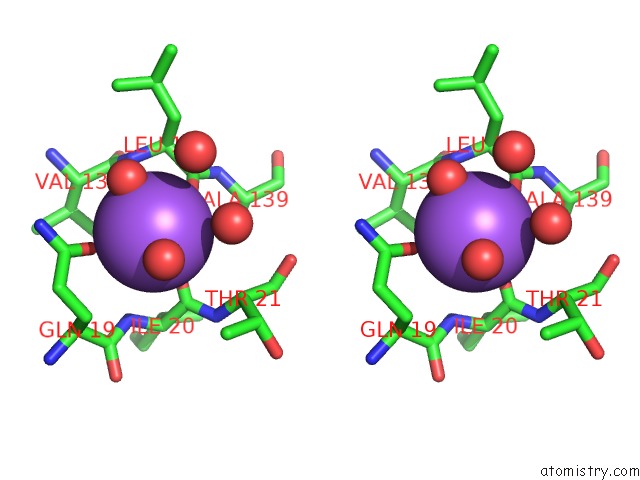
Stereo pair view
|
A full contact list of Sodium with other atoms in the Na binding
site number 1 of Crystal Structure of E. Coli Aminopeptidase N in Complex with L-Beta Homotryptophan within 5.0Å range:
probe
|
atom
|
residue
|
distance (Å)
|
B
|
Occ
|
A:Na903
b:21.7
occ:1.00
|
O
|
A:LEU138
|
2.3
|
14.7
|
1.0
|
O
|
A:HOH1549
|
2.4
|
17.5
|
1.0
|
O
|
A:HOH1753
|
2.4
|
29.4
|
1.0
|
O
|
A:ILE20
|
2.4
|
19.3
|
1.0
|
OE1
|
A:GLN19
|
2.5
|
18.5
|
1.0
|
C
|
A:LEU138
|
3.4
|
14.8
|
1.0
|
CD
|
A:GLN19
|
3.4
|
19.1
|
1.0
|
C
|
A:ILE20
|
3.5
|
16.6
|
1.0
|
N
|
A:LEU138
|
3.5
|
13.9
|
1.0
|
O
|
A:HOH1740
|
3.5
|
47.0
|
1.0
|
CA
|
A:LEU138
|
4.0
|
15.3
|
1.0
|
O
|
A:HOH1756
|
4.0
|
38.0
|
1.0
|
N
|
A:ILE20
|
4.0
|
16.4
|
1.0
|
CB
|
A:GLN19
|
4.1
|
16.4
|
1.0
|
CG
|
A:GLN19
|
4.2
|
19.1
|
1.0
|
NE2
|
A:GLN19
|
4.2
|
18.3
|
1.0
|
CA
|
A:THR21
|
4.2
|
16.1
|
1.0
|
CG1
|
A:VAL137
|
4.2
|
15.6
|
1.0
|
N
|
A:THR21
|
4.2
|
16.3
|
1.0
|
C
|
A:VAL137
|
4.3
|
14.1
|
1.0
|
CA
|
A:VAL137
|
4.4
|
14.9
|
1.0
|
N
|
A:ALA139
|
4.4
|
13.4
|
1.0
|
CA
|
A:ILE20
|
4.4
|
16.4
|
1.0
|
CB
|
A:LEU138
|
4.5
|
17.2
|
1.0
|
CA
|
A:GLN19
|
4.7
|
16.4
|
1.0
|
CA
|
A:ALA139
|
4.7
|
14.7
|
1.0
|
C
|
A:GLN19
|
4.8
|
18.8
|
1.0
|
CG2
|
A:THR21
|
4.8
|
21.1
|
1.0
|
CB
|
A:VAL137
|
5.0
|
14.2
|
1.0
|
|
Sodium binding site 2 out
of 8 in 4xnd
Go back to
Sodium Binding Sites List in 4xnd
Sodium binding site 2 out
of 8 in the Crystal Structure of E. Coli Aminopeptidase N in Complex with L-Beta Homotryptophan
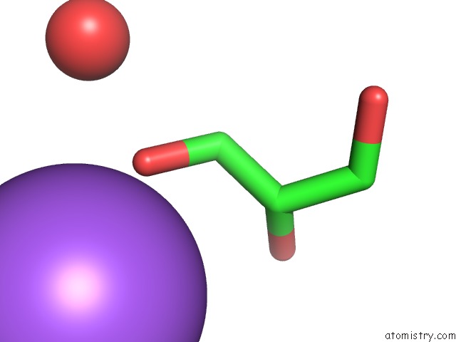
Mono view
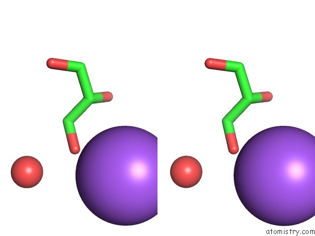
Stereo pair view
|
A full contact list of Sodium with other atoms in the Na binding
site number 2 of Crystal Structure of E. Coli Aminopeptidase N in Complex with L-Beta Homotryptophan within 5.0Å range:
probe
|
atom
|
residue
|
distance (Å)
|
B
|
Occ
|
A:Na904
b:30.3
occ:1.00
|
O1
|
A:GOL932
|
2.6
|
53.2
|
1.0
|
C1
|
A:GOL932
|
3.8
|
61.6
|
1.0
|
C2
|
A:GOL932
|
3.9
|
54.0
|
1.0
|
O2
|
A:GOL932
|
4.1
|
58.3
|
1.0
|
O
|
A:HOH1064
|
4.7
|
30.2
|
1.0
|
|
Sodium binding site 3 out
of 8 in 4xnd
Go back to
Sodium Binding Sites List in 4xnd
Sodium binding site 3 out
of 8 in the Crystal Structure of E. Coli Aminopeptidase N in Complex with L-Beta Homotryptophan
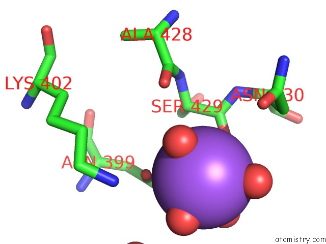
Mono view
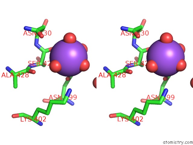
Stereo pair view
|
A full contact list of Sodium with other atoms in the Na binding
site number 3 of Crystal Structure of E. Coli Aminopeptidase N in Complex with L-Beta Homotryptophan within 5.0Å range:
probe
|
atom
|
residue
|
distance (Å)
|
B
|
Occ
|
A:Na905
b:53.5
occ:1.00
|
O
|
A:HOH1665
|
2.1
|
55.8
|
1.0
|
O
|
A:HOH1419
|
2.3
|
34.7
|
1.0
|
O
|
A:HOH1694
|
2.4
|
51.4
|
1.0
|
O
|
A:HOH1552
|
2.5
|
43.7
|
1.0
|
O
|
A:SER429
|
2.6
|
25.3
|
1.0
|
OD1
|
A:ASN399
|
2.8
|
28.1
|
1.0
|
ND2
|
A:ASN399
|
3.4
|
32.3
|
1.0
|
CG
|
A:ASN399
|
3.5
|
29.0
|
1.0
|
C
|
A:SER429
|
3.5
|
24.9
|
1.0
|
CA
|
A:SER429
|
3.9
|
22.1
|
1.0
|
NZ
|
A:LYS402
|
4.3
|
60.9
|
1.0
|
OD1
|
A:ASN430
|
4.4
|
49.6
|
1.0
|
O
|
A:ALA428
|
4.5
|
23.2
|
1.0
|
N
|
A:ASN430
|
4.5
|
22.2
|
1.0
|
CB
|
A:SER429
|
4.7
|
19.5
|
1.0
|
CB
|
A:ASN430
|
4.7
|
33.2
|
1.0
|
CB
|
A:ASN399
|
4.9
|
21.6
|
1.0
|
O
|
A:HOH1758
|
4.9
|
54.4
|
1.0
|
CG
|
A:ASN430
|
5.0
|
38.1
|
1.0
|
|
Sodium binding site 4 out
of 8 in 4xnd
Go back to
Sodium Binding Sites List in 4xnd
Sodium binding site 4 out
of 8 in the Crystal Structure of E. Coli Aminopeptidase N in Complex with L-Beta Homotryptophan
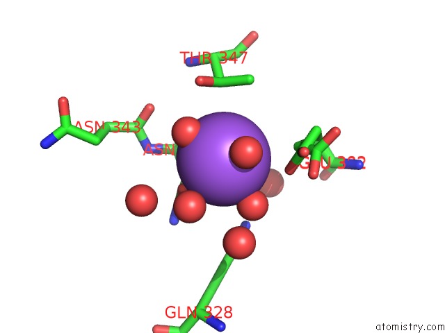
Mono view
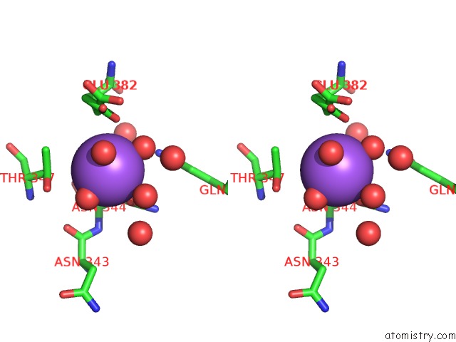
Stereo pair view
|
A full contact list of Sodium with other atoms in the Na binding
site number 4 of Crystal Structure of E. Coli Aminopeptidase N in Complex with L-Beta Homotryptophan within 5.0Å range:
probe
|
atom
|
residue
|
distance (Å)
|
B
|
Occ
|
A:Na906
b:39.4
occ:1.00
|
O
|
A:HOH1161
|
2.3
|
48.6
|
1.0
|
O
|
A:HOH1339
|
2.5
|
49.3
|
1.0
|
O
|
A:HOH1079
|
2.6
|
27.2
|
1.0
|
OD1
|
A:ASN344
|
2.7
|
19.7
|
1.0
|
O
|
A:HOH1344
|
2.8
|
19.1
|
1.0
|
OE1
|
A:GLU382
|
3.3
|
29.9
|
0.8
|
O
|
A:HOH1575
|
3.3
|
31.8
|
1.0
|
CG
|
A:ASN344
|
3.6
|
16.6
|
1.0
|
CD
|
A:GLU382
|
3.7
|
27.5
|
0.8
|
CG
|
A:GLU382
|
3.8
|
9.6
|
0.2
|
CA
|
A:ASN344
|
3.9
|
15.7
|
1.0
|
CB
|
A:THR347
|
3.9
|
17.4
|
1.0
|
OG1
|
A:THR347
|
3.9
|
16.5
|
1.0
|
CB
|
A:ASN344
|
3.9
|
16.8
|
1.0
|
OE2
|
A:GLU382
|
4.0
|
33.1
|
0.8
|
CG2
|
A:THR347
|
4.0
|
18.8
|
1.0
|
NE2
|
A:GLN328
|
4.2
|
14.6
|
1.0
|
O
|
A:HOH1047
|
4.4
|
29.0
|
1.0
|
OE1
|
A:GLU382
|
4.4
|
8.0
|
0.2
|
O
|
A:HOH1376
|
4.4
|
47.6
|
1.0
|
CD
|
A:GLU382
|
4.5
|
9.3
|
0.2
|
O
|
A:ASN344
|
4.6
|
13.9
|
1.0
|
ND2
|
A:ASN344
|
4.6
|
16.5
|
1.0
|
CG
|
A:GLU382
|
4.7
|
26.3
|
0.8
|
N
|
A:ASN344
|
4.8
|
15.0
|
1.0
|
C
|
A:ASN344
|
4.8
|
15.1
|
1.0
|
O
|
A:ASN343
|
4.8
|
14.9
|
1.0
|
O
|
A:HOH1166
|
5.0
|
14.1
|
1.0
|
|
Sodium binding site 5 out
of 8 in 4xnd
Go back to
Sodium Binding Sites List in 4xnd
Sodium binding site 5 out
of 8 in the Crystal Structure of E. Coli Aminopeptidase N in Complex with L-Beta Homotryptophan
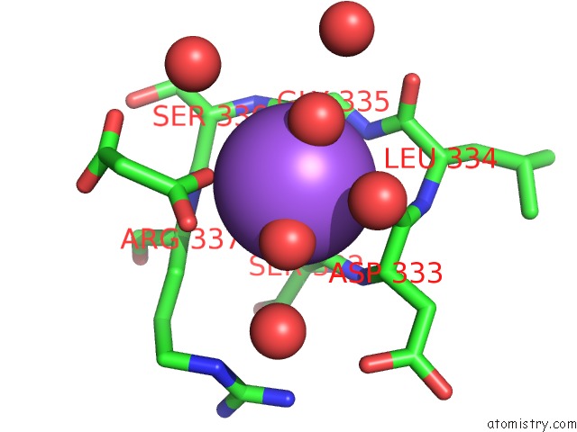
Mono view
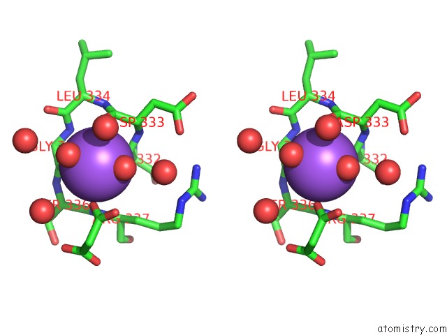
Stereo pair view
|
A full contact list of Sodium with other atoms in the Na binding
site number 5 of Crystal Structure of E. Coli Aminopeptidase N in Complex with L-Beta Homotryptophan within 5.0Å range:
probe
|
atom
|
residue
|
distance (Å)
|
B
|
Occ
|
A:Na907
b:22.0
occ:1.00
|
O
|
A:HOH1177
|
2.3
|
29.4
|
1.0
|
O
|
A:GLY335
|
2.3
|
16.9
|
1.0
|
O
|
A:HOH1648
|
2.4
|
27.4
|
1.0
|
O
|
A:SER332
|
2.4
|
17.6
|
1.0
|
O9
|
A:MLI936
|
2.5
|
25.1
|
1.0
|
O
|
A:ASP333
|
2.7
|
16.6
|
1.0
|
C
|
A:ASP333
|
3.4
|
18.1
|
1.0
|
C
|
A:GLY335
|
3.4
|
17.1
|
1.0
|
O
|
A:HOH1139
|
3.4
|
53.2
|
1.0
|
C3
|
A:MLI936
|
3.4
|
34.1
|
1.0
|
C
|
A:SER332
|
3.5
|
19.2
|
1.0
|
O8
|
A:MLI936
|
3.6
|
54.0
|
1.0
|
CA
|
A:ASP333
|
3.7
|
17.7
|
1.0
|
N
|
A:GLY335
|
3.9
|
16.0
|
1.0
|
N
|
A:ARG337
|
3.9
|
17.1
|
1.0
|
N
|
A:ASP333
|
4.1
|
17.2
|
1.0
|
O
|
A:HOH1425
|
4.2
|
31.3
|
1.0
|
N
|
A:LEU334
|
4.2
|
16.3
|
1.0
|
C
|
A:LEU334
|
4.2
|
18.2
|
1.0
|
CA
|
A:GLY335
|
4.3
|
17.7
|
1.0
|
N
|
A:SER336
|
4.3
|
16.8
|
1.0
|
O
|
A:HOH1581
|
4.3
|
48.3
|
1.0
|
CA
|
A:SER336
|
4.4
|
17.4
|
1.0
|
C
|
A:SER336
|
4.5
|
17.7
|
1.0
|
O
|
A:HOH1497
|
4.5
|
48.4
|
1.0
|
CB
|
A:ARG337
|
4.6
|
16.5
|
1.0
|
CA
|
A:LEU334
|
4.7
|
17.4
|
1.0
|
CA
|
A:ARG337
|
4.7
|
18.2
|
1.0
|
O
|
A:LEU334
|
4.8
|
16.7
|
1.0
|
CA
|
A:SER332
|
4.8
|
16.7
|
1.0
|
C1
|
A:MLI936
|
4.8
|
38.2
|
1.0
|
|
Sodium binding site 6 out
of 8 in 4xnd
Go back to
Sodium Binding Sites List in 4xnd
Sodium binding site 6 out
of 8 in the Crystal Structure of E. Coli Aminopeptidase N in Complex with L-Beta Homotryptophan
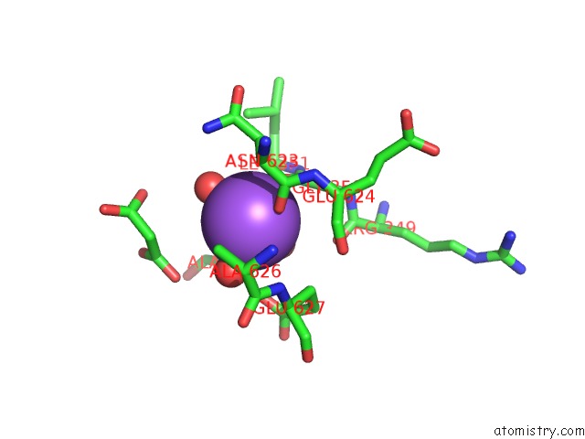
Mono view
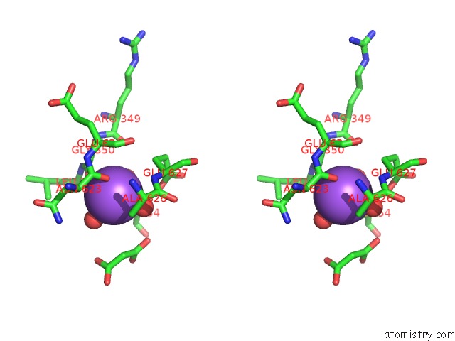
Stereo pair view
|
A full contact list of Sodium with other atoms in the Na binding
site number 6 of Crystal Structure of E. Coli Aminopeptidase N in Complex with L-Beta Homotryptophan within 5.0Å range:
probe
|
atom
|
residue
|
distance (Å)
|
B
|
Occ
|
A:Na908
b:32.0
occ:1.00
|
O
|
A:HOH1645
|
2.4
|
40.9
|
1.0
|
O
|
A:ASN623
|
2.4
|
20.6
|
1.0
|
O
|
A:HOH1028
|
2.4
|
28.9
|
1.0
|
O
|
A:GLY350
|
2.6
|
23.2
|
1.0
|
O
|
A:HOH1223
|
2.7
|
27.1
|
1.0
|
C
|
A:ASN623
|
3.4
|
23.6
|
1.0
|
C
|
A:GLY350
|
3.5
|
23.9
|
1.0
|
CB
|
A:ALA354
|
3.7
|
20.3
|
1.0
|
OE1
|
A:GLU627
|
3.9
|
32.1
|
1.0
|
CA
|
A:ASN623
|
3.9
|
25.2
|
1.0
|
CB
|
A:ASN623
|
4.0
|
29.7
|
1.0
|
CA
|
A:GLY350
|
4.0
|
21.3
|
1.0
|
N
|
A:GLU627
|
4.1
|
20.4
|
1.0
|
CB
|
A:GLU627
|
4.2
|
27.2
|
1.0
|
CB
|
A:ALA626
|
4.2
|
19.4
|
1.0
|
O9
|
A:MLI933
|
4.4
|
37.0
|
1.0
|
N
|
A:GLU624
|
4.4
|
21.3
|
1.0
|
CA
|
A:GLU627
|
4.5
|
22.3
|
1.0
|
N
|
A:LEU351
|
4.6
|
19.2
|
1.0
|
O
|
A:ARG349
|
4.6
|
22.6
|
1.0
|
C1
|
A:MLI933
|
4.6
|
38.6
|
1.0
|
C
|
A:ALA626
|
4.7
|
20.1
|
1.0
|
C3
|
A:MLI933
|
4.7
|
37.3
|
1.0
|
CA
|
A:GLU624
|
4.8
|
21.8
|
1.0
|
CA
|
A:LEU351
|
4.8
|
17.1
|
1.0
|
CA
|
A:ALA354
|
4.8
|
18.9
|
1.0
|
CD
|
A:GLU627
|
4.8
|
34.7
|
1.0
|
ND2
|
A:ASN623
|
4.9
|
34.3
|
1.0
|
CA
|
A:ALA626
|
4.9
|
20.5
|
1.0
|
N
|
A:ALA354
|
4.9
|
16.3
|
1.0
|
CG
|
A:ASN623
|
5.0
|
29.2
|
1.0
|
|
Sodium binding site 7 out
of 8 in 4xnd
Go back to
Sodium Binding Sites List in 4xnd
Sodium binding site 7 out
of 8 in the Crystal Structure of E. Coli Aminopeptidase N in Complex with L-Beta Homotryptophan
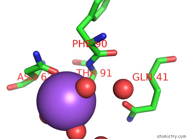
Mono view
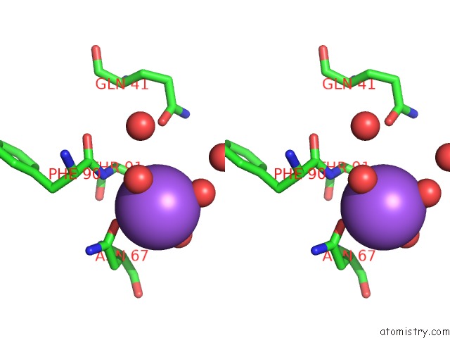
Stereo pair view
|
A full contact list of Sodium with other atoms in the Na binding
site number 7 of Crystal Structure of E. Coli Aminopeptidase N in Complex with L-Beta Homotryptophan within 5.0Å range:
probe
|
atom
|
residue
|
distance (Å)
|
B
|
Occ
|
A:Na909
b:43.4
occ:1.00
|
O
|
A:HOH1710
|
2.4
|
56.1
|
1.0
|
O
|
A:HOH1724
|
2.5
|
34.3
|
1.0
|
OD1
|
A:ASN67
|
2.5
|
20.5
|
1.0
|
O
|
A:HOH1745
|
2.6
|
40.6
|
1.0
|
OG1
|
A:THR91
|
2.7
|
19.2
|
1.0
|
CG
|
A:ASN67
|
3.4
|
24.8
|
1.0
|
CB
|
A:THR91
|
3.4
|
18.0
|
1.0
|
N
|
A:THR91
|
3.8
|
16.3
|
1.0
|
CB
|
A:ASN67
|
4.2
|
22.9
|
1.0
|
CA
|
A:THR91
|
4.2
|
18.1
|
1.0
|
ND2
|
A:ASN67
|
4.2
|
22.3
|
1.0
|
CA
|
A:ASN67
|
4.4
|
21.5
|
1.0
|
O
|
A:HOH1785
|
4.4
|
58.4
|
1.0
|
O
|
A:HOH1248
|
4.5
|
29.8
|
1.0
|
C
|
A:PHE90
|
4.5
|
16.8
|
1.0
|
CG2
|
A:THR91
|
4.6
|
21.0
|
1.0
|
CA
|
A:PHE90
|
4.7
|
18.4
|
1.0
|
OE1
|
A:GLN41
|
4.8
|
24.9
|
1.0
|
|
Sodium binding site 8 out
of 8 in 4xnd
Go back to
Sodium Binding Sites List in 4xnd
Sodium binding site 8 out
of 8 in the Crystal Structure of E. Coli Aminopeptidase N in Complex with L-Beta Homotryptophan
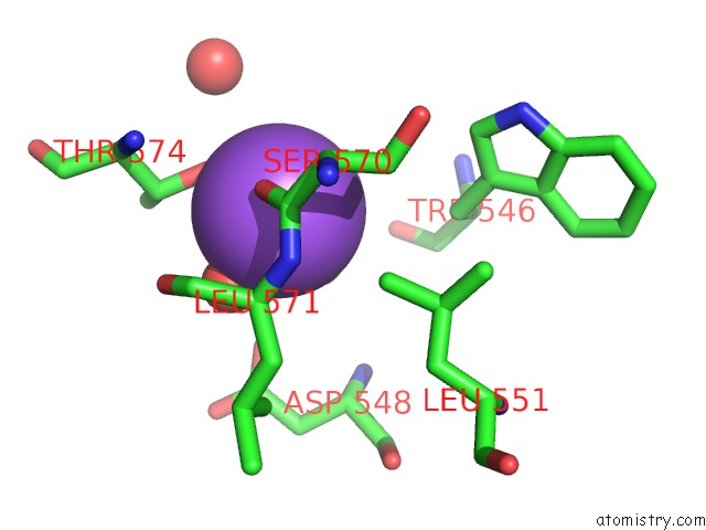
Mono view
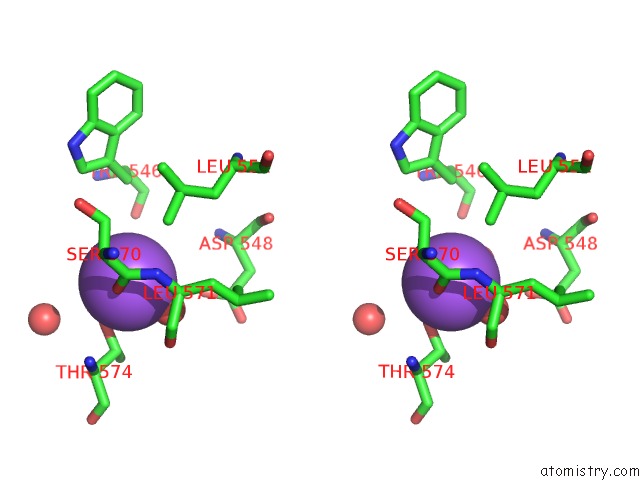
Stereo pair view
|
A full contact list of Sodium with other atoms in the Na binding
site number 8 of Crystal Structure of E. Coli Aminopeptidase N in Complex with L-Beta Homotryptophan within 5.0Å range:
probe
|
atom
|
residue
|
distance (Å)
|
B
|
Occ
|
A:Na910
b:33.5
occ:1.00
|
OG1
|
A:THR574
|
2.3
|
19.6
|
1.0
|
O
|
A:HOH1328
|
2.3
|
31.4
|
1.0
|
O
|
A:HOH1390
|
2.4
|
27.3
|
1.0
|
O
|
A:SER570
|
2.6
|
20.8
|
1.0
|
CB
|
A:THR574
|
3.3
|
21.4
|
1.0
|
C
|
A:SER570
|
3.6
|
19.4
|
1.0
|
CD1
|
A:LEU551
|
3.6
|
17.3
|
1.0
|
CA
|
A:LEU571
|
3.9
|
18.5
|
1.0
|
O
|
A:LEU571
|
4.1
|
22.7
|
1.0
|
N
|
A:LEU571
|
4.1
|
17.8
|
1.0
|
CG2
|
A:THR574
|
4.2
|
25.1
|
1.0
|
N
|
A:THR574
|
4.2
|
17.6
|
1.0
|
OD1
|
A:ASP548
|
4.2
|
24.4
|
1.0
|
C
|
A:LEU571
|
4.3
|
20.0
|
1.0
|
CA
|
A:THR574
|
4.4
|
19.0
|
1.0
|
O
|
A:HOH1500
|
4.5
|
41.9
|
1.0
|
CA
|
A:SER570
|
4.7
|
16.5
|
1.0
|
CB
|
A:SER570
|
4.8
|
18.0
|
1.0
|
CB
|
A:TRP546
|
5.0
|
24.9
|
1.0
|
|
Reference:
A.Addlagatta,
R.Gumpena.
Crystal Structure of E. Coli Aminopeptidase N in Complex with L-Beta Homotryptophan To Be Published.
Page generated: Mon Oct 7 19:15:47 2024
|