Sodium »
PDB 4qon-4r3n »
4qpk »
Sodium in PDB 4qpk: 1.7 Angstrom Structure of A Bacterial Phosphotransferase
Protein crystallography data
The structure of 1.7 Angstrom Structure of A Bacterial Phosphotransferase, PDB code: 4qpk
was solved by
J.W.Willett,
S.Crosson,
J.Herrou,
with X-Ray Crystallography technique. A brief refinement statistics is given in the table below:
| Resolution Low / High (Å) | 35.42 / 1.66 |
| Space group | P 43 |
| Cell size a, b, c (Å), α, β, γ (°) | 70.840, 70.840, 87.010, 90.00, 90.00, 90.00 |
| R / Rfree (%) | 16.4 / 18.8 |
Sodium Binding Sites:
The binding sites of Sodium atom in the 1.7 Angstrom Structure of A Bacterial Phosphotransferase
(pdb code 4qpk). This binding sites where shown within
5.0 Angstroms radius around Sodium atom.
In total only one binding site of Sodium was determined in the 1.7 Angstrom Structure of A Bacterial Phosphotransferase, PDB code: 4qpk:
In total only one binding site of Sodium was determined in the 1.7 Angstrom Structure of A Bacterial Phosphotransferase, PDB code: 4qpk:
Sodium binding site 1 out of 1 in 4qpk
Go back to
Sodium binding site 1 out
of 1 in the 1.7 Angstrom Structure of A Bacterial Phosphotransferase
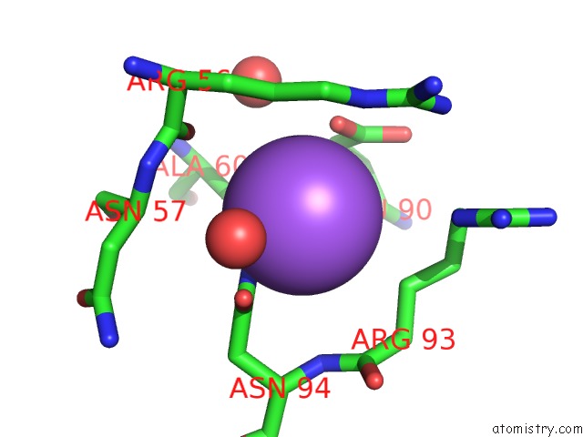
Mono view
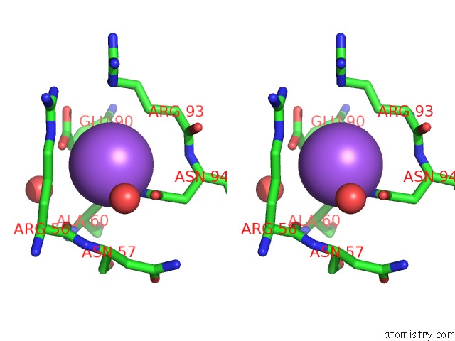
Stereo pair view
Mono view
Stereo pair view
A full contact list of Sodium with other atoms in the Na binding
site number 1 of 1.7 Angstrom Structure of A Bacterial Phosphotransferase within 5.0Å range:
|
Reference:
J.W.Willett,
J.Herrou,
A.Briegel,
G.Rotskoff,
S.Crosson.
Structural Asymmetry in A Conserved Signaling System That Regulates Division, Replication, and Virulence of An Intracellular Pathogen. Proc.Natl.Acad.Sci.Usa V. 112 E3709 2015.
ISSN: ISSN 0027-8424
PubMed: 26124143
DOI: 10.1073/PNAS.1503118112
Page generated: Mon Oct 7 18:05:20 2024
ISSN: ISSN 0027-8424
PubMed: 26124143
DOI: 10.1073/PNAS.1503118112
Last articles
Zn in 9J0NZn in 9J0O
Zn in 9J0P
Zn in 9FJX
Zn in 9EKB
Zn in 9C0F
Zn in 9CAH
Zn in 9CH0
Zn in 9CH3
Zn in 9CH1