Sodium »
PDB 4owe-4pd7 »
4p4w »
Sodium in PDB 4p4w: Dodecamer Formed By A Macrocyclic Peptide Derived From Beta-2- Microglobulin (63-69) - (Orn)Yll(Phi)Yte(Orn)Kva(Mva)Avk
Protein crystallography data
The structure of Dodecamer Formed By A Macrocyclic Peptide Derived From Beta-2- Microglobulin (63-69) - (Orn)Yll(Phi)Yte(Orn)Kva(Mva)Avk, PDB code: 4p4w
was solved by
R.K.Spencer,
A.Kreutzer,
J.S.Nowick,
with X-Ray Crystallography technique. A brief refinement statistics is given in the table below:
| Resolution Low / High (Å) | 31.53 / 1.50 |
| Space group | P 31 2 1 |
| Cell size a, b, c (Å), α, β, γ (°) | 58.193, 58.193, 121.279, 90.00, 90.00, 120.00 |
| R / Rfree (%) | 17.5 / 20.9 |
Other elements in 4p4w:
The structure of Dodecamer Formed By A Macrocyclic Peptide Derived From Beta-2- Microglobulin (63-69) - (Orn)Yll(Phi)Yte(Orn)Kva(Mva)Avk also contains other interesting chemical elements:
| Iodine | (I) | 14 atoms |
| Chlorine | (Cl) | 6 atoms |
Sodium Binding Sites:
The binding sites of Sodium atom in the Dodecamer Formed By A Macrocyclic Peptide Derived From Beta-2- Microglobulin (63-69) - (Orn)Yll(Phi)Yte(Orn)Kva(Mva)Avk
(pdb code 4p4w). This binding sites where shown within
5.0 Angstroms radius around Sodium atom.
In total only one binding site of Sodium was determined in the Dodecamer Formed By A Macrocyclic Peptide Derived From Beta-2- Microglobulin (63-69) - (Orn)Yll(Phi)Yte(Orn)Kva(Mva)Avk, PDB code: 4p4w:
In total only one binding site of Sodium was determined in the Dodecamer Formed By A Macrocyclic Peptide Derived From Beta-2- Microglobulin (63-69) - (Orn)Yll(Phi)Yte(Orn)Kva(Mva)Avk, PDB code: 4p4w:
Sodium binding site 1 out of 1 in 4p4w
Go back to
Sodium binding site 1 out
of 1 in the Dodecamer Formed By A Macrocyclic Peptide Derived From Beta-2- Microglobulin (63-69) - (Orn)Yll(Phi)Yte(Orn)Kva(Mva)Avk
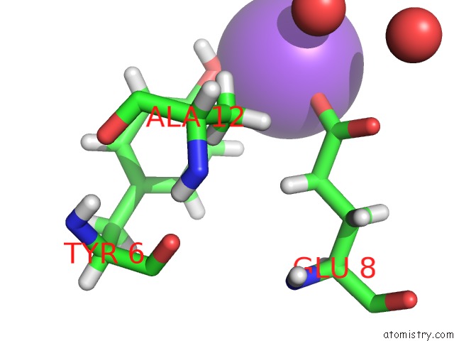
Mono view
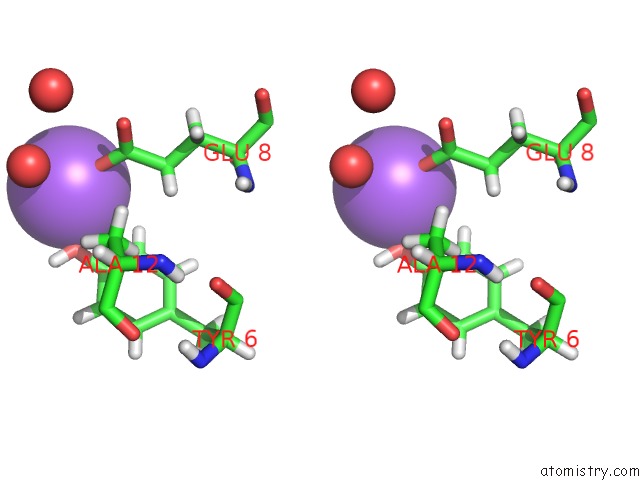
Stereo pair view
Mono view
Stereo pair view
A full contact list of Sodium with other atoms in the Na binding
site number 1 of Dodecamer Formed By A Macrocyclic Peptide Derived From Beta-2- Microglobulin (63-69) - (Orn)Yll(Phi)Yte(Orn)Kva(Mva)Avk within 5.0Å range:
|
Reference:
R.K.Spencer,
A.G.Kreutzer,
P.J.Salveson,
H.Li,
J.S.Nowick.
X-Ray Crystallographic Structures of Oligomers of Peptides Derived From Beta 2-Microglobulin. J.Am.Chem.Soc. 2015.
ISSN: ESSN 1520-5126
PubMed: 25915729
DOI: 10.1021/JACS.5B01673
Page generated: Mon Oct 7 17:38:27 2024
ISSN: ESSN 1520-5126
PubMed: 25915729
DOI: 10.1021/JACS.5B01673
Last articles
Ca in 2Y7ZCa in 2Y73
Ca in 2Y7X
Ca in 2Y79
Ca in 2Y6K
Ca in 2Y6L
Ca in 2Y6J
Ca in 2Y5I
Ca in 2Y6H
Ca in 2Y6G