Sodium »
PDB 4oci-4owc »
4oki »
Sodium in PDB 4oki: X-Ray Structure of the Nucleotide-Binding Subdomain of the Enoylreductase Domain of Ppsc From Mycobacterium Tuberculosis
Enzymatic activity of X-Ray Structure of the Nucleotide-Binding Subdomain of the Enoylreductase Domain of Ppsc From Mycobacterium Tuberculosis
All present enzymatic activity of X-Ray Structure of the Nucleotide-Binding Subdomain of the Enoylreductase Domain of Ppsc From Mycobacterium Tuberculosis:
2.3.1.41;
2.3.1.41;
Protein crystallography data
The structure of X-Ray Structure of the Nucleotide-Binding Subdomain of the Enoylreductase Domain of Ppsc From Mycobacterium Tuberculosis, PDB code: 4oki
was solved by
A.Faille,
L.Mourey,
J.D.Pedelacq,
with X-Ray Crystallography technique. A brief refinement statistics is given in the table below:
| Resolution Low / High (Å) | 42.00 / 1.50 |
| Space group | P 21 21 21 |
| Cell size a, b, c (Å), α, β, γ (°) | 59.885, 78.743, 88.796, 90.00, 90.00, 90.00 |
| R / Rfree (%) | 18.2 / 21.8 |
Sodium Binding Sites:
The binding sites of Sodium atom in the X-Ray Structure of the Nucleotide-Binding Subdomain of the Enoylreductase Domain of Ppsc From Mycobacterium Tuberculosis
(pdb code 4oki). This binding sites where shown within
5.0 Angstroms radius around Sodium atom.
In total 7 binding sites of Sodium where determined in the X-Ray Structure of the Nucleotide-Binding Subdomain of the Enoylreductase Domain of Ppsc From Mycobacterium Tuberculosis, PDB code: 4oki:
Jump to Sodium binding site number: 1; 2; 3; 4; 5; 6; 7;
In total 7 binding sites of Sodium where determined in the X-Ray Structure of the Nucleotide-Binding Subdomain of the Enoylreductase Domain of Ppsc From Mycobacterium Tuberculosis, PDB code: 4oki:
Jump to Sodium binding site number: 1; 2; 3; 4; 5; 6; 7;
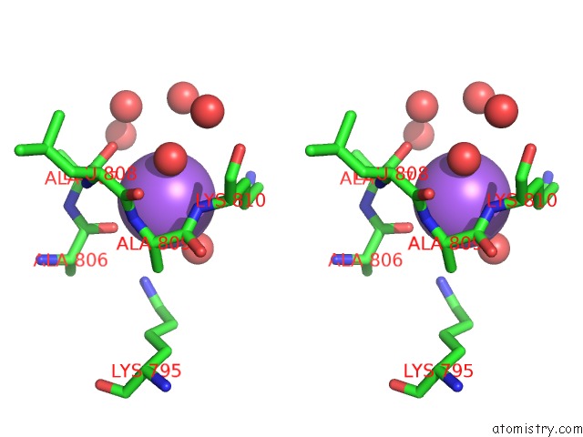
Sodium binding site 1 out of 7 in 4oki
Go back to
Sodium binding site 1 out
of 7 in the X-Ray Structure of the Nucleotide-Binding Subdomain of the Enoylreductase Domain of Ppsc From Mycobacterium Tuberculosis
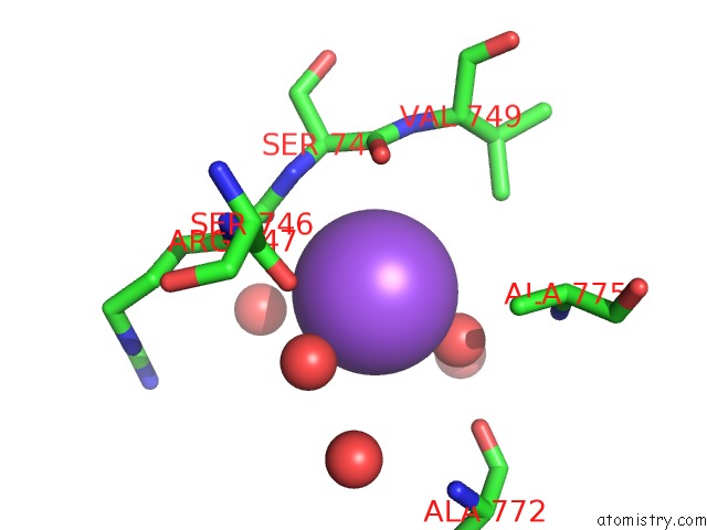
Mono view
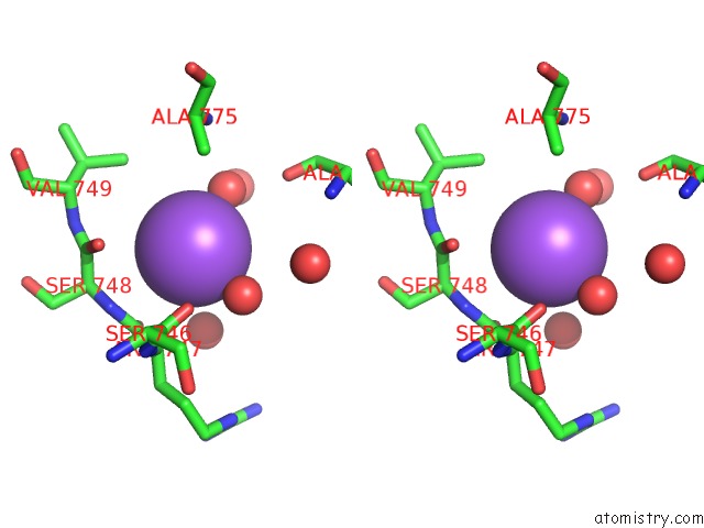
Stereo pair view
Mono view
Stereo pair view
A full contact list of Sodium with other atoms in the Na binding
site number 1 of X-Ray Structure of the Nucleotide-Binding Subdomain of the Enoylreductase Domain of Ppsc From Mycobacterium Tuberculosis within 5.0Å range:
|
Sodium binding site 2 out of 7 in 4oki
Go back to
Sodium binding site 2 out
of 7 in the X-Ray Structure of the Nucleotide-Binding Subdomain of the Enoylreductase Domain of Ppsc From Mycobacterium Tuberculosis
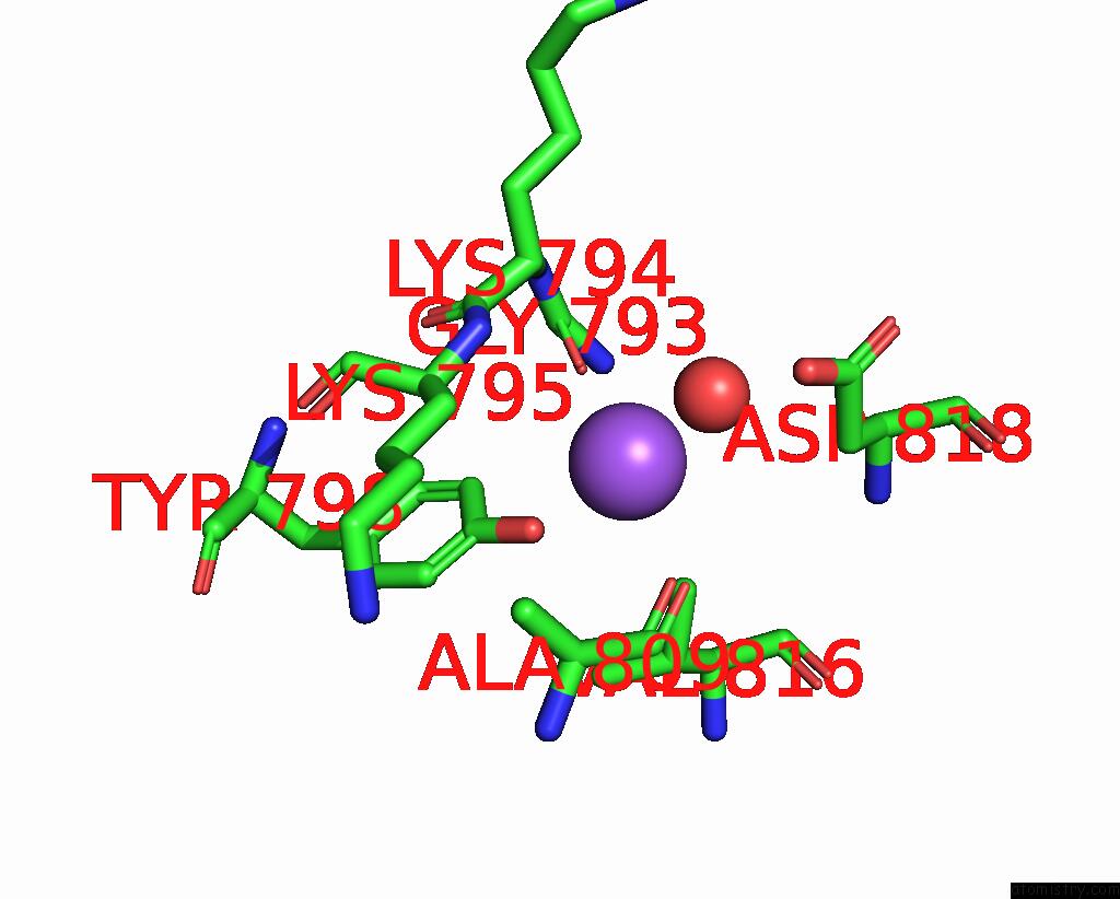
Mono view

Stereo pair view
Mono view
Stereo pair view
A full contact list of Sodium with other atoms in the Na binding
site number 2 of X-Ray Structure of the Nucleotide-Binding Subdomain of the Enoylreductase Domain of Ppsc From Mycobacterium Tuberculosis within 5.0Å range:
|
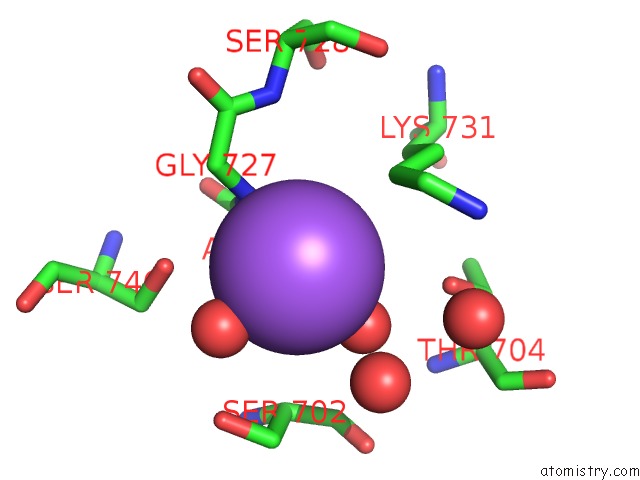
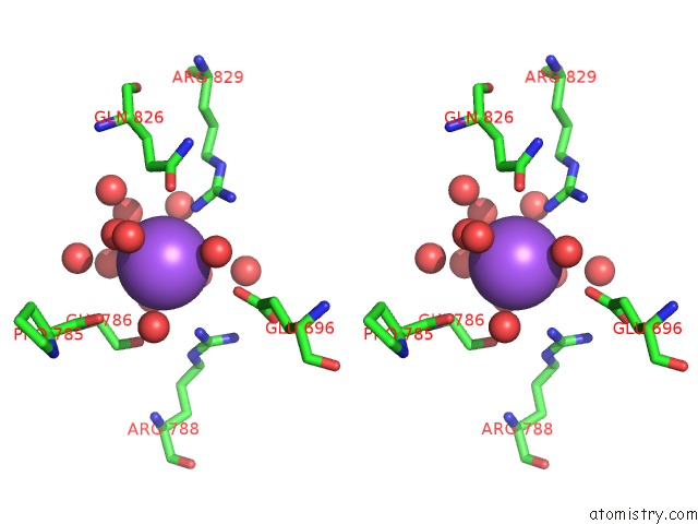
Sodium binding site 3 out of 7 in 4oki
Go back to
Sodium binding site 3 out
of 7 in the X-Ray Structure of the Nucleotide-Binding Subdomain of the Enoylreductase Domain of Ppsc From Mycobacterium Tuberculosis

Mono view

Stereo pair view
Mono view
Stereo pair view
A full contact list of Sodium with other atoms in the Na binding
site number 3 of X-Ray Structure of the Nucleotide-Binding Subdomain of the Enoylreductase Domain of Ppsc From Mycobacterium Tuberculosis within 5.0Å range:
|
Sodium binding site 4 out of 7 in 4oki
Go back to
Sodium binding site 4 out
of 7 in the X-Ray Structure of the Nucleotide-Binding Subdomain of the Enoylreductase Domain of Ppsc From Mycobacterium Tuberculosis
Mono view

Stereo pair view
Mono view
Stereo pair view
A full contact list of Sodium with other atoms in the Na binding
site number 4 of X-Ray Structure of the Nucleotide-Binding Subdomain of the Enoylreductase Domain of Ppsc From Mycobacterium Tuberculosis within 5.0Å range:
|
Sodium binding site 5 out of 7 in 4oki
Go back to
Sodium binding site 5 out
of 7 in the X-Ray Structure of the Nucleotide-Binding Subdomain of the Enoylreductase Domain of Ppsc From Mycobacterium Tuberculosis

Mono view
Stereo pair view
Mono view
Stereo pair view
A full contact list of Sodium with other atoms in the Na binding
site number 5 of X-Ray Structure of the Nucleotide-Binding Subdomain of the Enoylreductase Domain of Ppsc From Mycobacterium Tuberculosis within 5.0Å range:
|
Sodium binding site 6 out of 7 in 4oki
Go back to
Sodium binding site 6 out
of 7 in the X-Ray Structure of the Nucleotide-Binding Subdomain of the Enoylreductase Domain of Ppsc From Mycobacterium Tuberculosis

Mono view
Stereo pair view
Mono view
Stereo pair view
A full contact list of Sodium with other atoms in the Na binding
site number 6 of X-Ray Structure of the Nucleotide-Binding Subdomain of the Enoylreductase Domain of Ppsc From Mycobacterium Tuberculosis within 5.0Å range:
|
Sodium binding site 7 out of 7 in 4oki
Go back to
Sodium binding site 7 out
of 7 in the X-Ray Structure of the Nucleotide-Binding Subdomain of the Enoylreductase Domain of Ppsc From Mycobacterium Tuberculosis
Mono view

Stereo pair view
Mono view
Stereo pair view
A full contact list of Sodium with other atoms in the Na binding
site number 7 of X-Ray Structure of the Nucleotide-Binding Subdomain of the Enoylreductase Domain of Ppsc From Mycobacterium Tuberculosis within 5.0Å range:
|
Reference:
A.Faille,
N.Slama,
A.Quemard,
L.Mourey,
J.D.Pedelacq.
Insights Into the Catalytic Mechanism of the Dh Domain of the Mycobacterium Tuberculosis Polyketide Synthase Ppsc and Architecture of the Beta-Carbon Processing Domains To Be Published.
Page generated: Mon Oct 7 17:29:09 2024
Last articles
Cl in 7TIVCl in 7TIW
Cl in 7TI9
Cl in 7TIU
Cl in 7TIN
Cl in 7TI7
Cl in 7TI1
Cl in 7TI0
Cl in 7TH2
Cl in 7TGF