Sodium »
PDB 4mfc-4mzx »
4mlm »
Sodium in PDB 4mlm: Crystal Structure of Phnz From Uncultured Bacterium HF130_AEPN_1
Protein crystallography data
The structure of Crystal Structure of Phnz From Uncultured Bacterium HF130_AEPN_1, PDB code: 4mlm
was solved by
L.M.Van Staalduinen,
F.R.Mcsorley,
D.L.Zechel,
Z.Jia,
Montreal-Kingstonbacterial Structural Genomics Initiative (Bsgi),
with X-Ray Crystallography technique. A brief refinement statistics is given in the table below:
| Resolution Low / High (Å) | 19.80 / 1.70 |
| Space group | P 21 21 2 |
| Cell size a, b, c (Å), α, β, γ (°) | 104.670, 76.040, 60.700, 90.00, 90.00, 90.00 |
| R / Rfree (%) | 17.2 / 19.8 |
Other elements in 4mlm:
The structure of Crystal Structure of Phnz From Uncultured Bacterium HF130_AEPN_1 also contains other interesting chemical elements:
| Iron | (Fe) | 4 atoms |
Sodium Binding Sites:
The binding sites of Sodium atom in the Crystal Structure of Phnz From Uncultured Bacterium HF130_AEPN_1
(pdb code 4mlm). This binding sites where shown within
5.0 Angstroms radius around Sodium atom.
In total only one binding site of Sodium was determined in the Crystal Structure of Phnz From Uncultured Bacterium HF130_AEPN_1, PDB code: 4mlm:
In total only one binding site of Sodium was determined in the Crystal Structure of Phnz From Uncultured Bacterium HF130_AEPN_1, PDB code: 4mlm:
Sodium binding site 1 out of 1 in 4mlm
Go back to
Sodium binding site 1 out
of 1 in the Crystal Structure of Phnz From Uncultured Bacterium HF130_AEPN_1
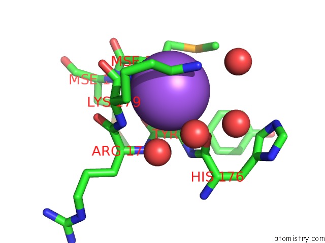
Mono view
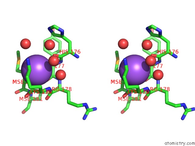
Stereo pair view
Mono view
Stereo pair view
A full contact list of Sodium with other atoms in the Na binding
site number 1 of Crystal Structure of Phnz From Uncultured Bacterium HF130_AEPN_1 within 5.0Å range:
|
Reference:
L.M.Van Staalduinen,
F.R.Mcsorley,
K.Schiessl,
J.Seguin,
P.B.Wyatt,
F.Hammerschmidt,
D.L.Zechel,
Z.Jia.
Crystal Structure of Phnz in Complex with Substrate Reveals A Di-Iron Oxygenase Mechanism For Catabolism of Organophosphonates. Proc.Natl.Acad.Sci.Usa V. 111 5171 2014.
ISSN: ISSN 0027-8424
PubMed: 24706911
DOI: 10.1073/PNAS.1320039111
Page generated: Mon Oct 7 17:00:22 2024
ISSN: ISSN 0027-8424
PubMed: 24706911
DOI: 10.1073/PNAS.1320039111
Last articles
Zn in 9MJ5Zn in 9HNW
Zn in 9G0L
Zn in 9FNE
Zn in 9DZN
Zn in 9E0I
Zn in 9D32
Zn in 9DAK
Zn in 8ZXC
Zn in 8ZUF