Sodium »
PDB 4m04-4mfa »
4mb6 »
Sodium in PDB 4mb6: Crystal Structure of Adenine Phosphoribosyltransferase From Yersinia Pseudotuberculosis.
Enzymatic activity of Crystal Structure of Adenine Phosphoribosyltransferase From Yersinia Pseudotuberculosis.
All present enzymatic activity of Crystal Structure of Adenine Phosphoribosyltransferase From Yersinia Pseudotuberculosis.:
2.4.2.7;
2.4.2.7;
Protein crystallography data
The structure of Crystal Structure of Adenine Phosphoribosyltransferase From Yersinia Pseudotuberculosis., PDB code: 4mb6
was solved by
G.C.Pavithra,
J.Kim,
R.P.Hegde,
S.C.Almo,
U.A.Ramagopal,
New Yorkstructural Genomics Research Consortium (Nysgrc),
with X-Ray Crystallography technique. A brief refinement statistics is given in the table below:
| Resolution Low / High (Å) | 40.19 / 1.81 |
| Space group | C 1 2 1 |
| Cell size a, b, c (Å), α, β, γ (°) | 58.560, 78.380, 54.010, 90.00, 112.98, 90.00 |
| R / Rfree (%) | 18.4 / 23.8 |
Sodium Binding Sites:
The binding sites of Sodium atom in the Crystal Structure of Adenine Phosphoribosyltransferase From Yersinia Pseudotuberculosis.
(pdb code 4mb6). This binding sites where shown within
5.0 Angstroms radius around Sodium atom.
In total only one binding site of Sodium was determined in the Crystal Structure of Adenine Phosphoribosyltransferase From Yersinia Pseudotuberculosis., PDB code: 4mb6:
In total only one binding site of Sodium was determined in the Crystal Structure of Adenine Phosphoribosyltransferase From Yersinia Pseudotuberculosis., PDB code: 4mb6:
Sodium binding site 1 out of 1 in 4mb6
Go back to
Sodium binding site 1 out
of 1 in the Crystal Structure of Adenine Phosphoribosyltransferase From Yersinia Pseudotuberculosis.
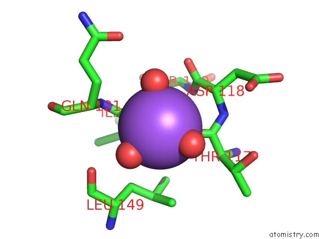
Mono view
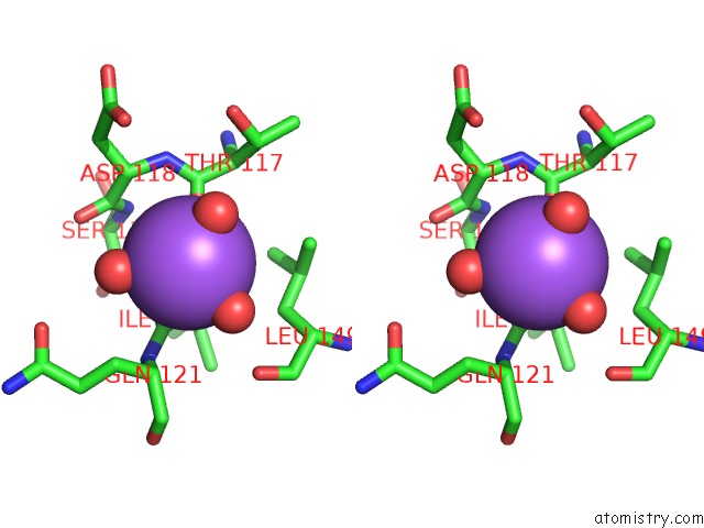
Stereo pair view
Mono view
Stereo pair view
A full contact list of Sodium with other atoms in the Na binding
site number 1 of Crystal Structure of Adenine Phosphoribosyltransferase From Yersinia Pseudotuberculosis. within 5.0Å range:
|
Reference:
G.C.Pavithra,
J.Kim,
R.P.Hegde,
S.C.Almo,
U.A.Ramagopal.
Crystal Structure of Adenine Phosphoribosyltransferase From Yersinia Pseudotuberculosis To Be Published.
Page generated: Mon Oct 7 16:58:58 2024
Last articles
Cl in 7YBPCl in 7YBO
Cl in 7Y94
Cl in 7Y6I
Cl in 7YBG
Cl in 7Y7Z
Cl in 7Y8I
Cl in 7Y7W
Cl in 7Y7Y
Cl in 7Y7V