Sodium »
PDB 4m04-4mfa »
4mb5 »
Sodium in PDB 4mb5: Crystal Structure of E153Q Mutant of Cold-Adapted Chitinase From Moritella Complex with NAG5
Enzymatic activity of Crystal Structure of E153Q Mutant of Cold-Adapted Chitinase From Moritella Complex with NAG5
All present enzymatic activity of Crystal Structure of E153Q Mutant of Cold-Adapted Chitinase From Moritella Complex with NAG5:
3.2.1.14;
3.2.1.14;
Protein crystallography data
The structure of Crystal Structure of E153Q Mutant of Cold-Adapted Chitinase From Moritella Complex with NAG5, PDB code: 4mb5
was solved by
P.H.Malecki,
C.E.Vorgias,
W.Rypniewski,
with X-Ray Crystallography technique. A brief refinement statistics is given in the table below:
| Resolution Low / High (Å) | 34.44 / 1.64 |
| Space group | P 31 1 2 |
| Cell size a, b, c (Å), α, β, γ (°) | 67.057, 67.057, 256.633, 90.00, 90.00, 120.00 |
| R / Rfree (%) | 14.6 / 17.7 |
Sodium Binding Sites:
The binding sites of Sodium atom in the Crystal Structure of E153Q Mutant of Cold-Adapted Chitinase From Moritella Complex with NAG5
(pdb code 4mb5). This binding sites where shown within
5.0 Angstroms radius around Sodium atom.
In total only one binding site of Sodium was determined in the Crystal Structure of E153Q Mutant of Cold-Adapted Chitinase From Moritella Complex with NAG5, PDB code: 4mb5:
In total only one binding site of Sodium was determined in the Crystal Structure of E153Q Mutant of Cold-Adapted Chitinase From Moritella Complex with NAG5, PDB code: 4mb5:
Sodium binding site 1 out of 1 in 4mb5
Go back to
Sodium binding site 1 out
of 1 in the Crystal Structure of E153Q Mutant of Cold-Adapted Chitinase From Moritella Complex with NAG5
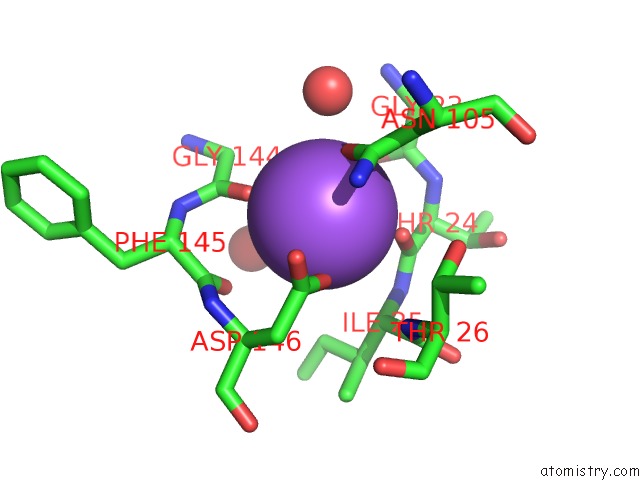
Mono view
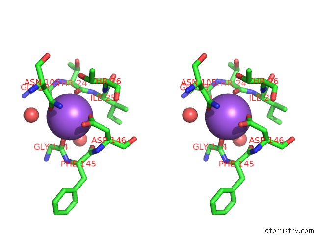
Stereo pair view
Mono view
Stereo pair view
A full contact list of Sodium with other atoms in the Na binding
site number 1 of Crystal Structure of E153Q Mutant of Cold-Adapted Chitinase From Moritella Complex with NAG5 within 5.0Å range:
|
Reference:
P.H.Malecki,
C.E.Vorgias,
M.V.Petoukhov,
D.I.Svergun,
W.Rypniewski.
Crystal Structures of Substrate-Bound Chitinase From the Psychrophilic Bacterium Moritella Marina and Its Structure in Solution Acta Crystallogr.,Sect.D V. 70 676 2014.
ISSN: ISSN 0907-4449
PubMed: 24598737
DOI: 10.1107/S1399004713032264
Page generated: Mon Oct 7 16:58:58 2024
ISSN: ISSN 0907-4449
PubMed: 24598737
DOI: 10.1107/S1399004713032264
Last articles
Zn in 9MJ5Zn in 9HNW
Zn in 9G0L
Zn in 9FNE
Zn in 9DZN
Zn in 9E0I
Zn in 9D32
Zn in 9DAK
Zn in 8ZXC
Zn in 8ZUF