Sodium »
PDB 4hfd-4i05 »
4hu7 »
Sodium in PDB 4hu7: E. Coli Thioredoxin Variant with PRO76 As Single Proline Residue
Protein crystallography data
The structure of E. Coli Thioredoxin Variant with PRO76 As Single Proline Residue, PDB code: 4hu7
was solved by
R.Glockshuber,
M.A.Scharer,
G.Capitani,
M.Rubini,
with X-Ray Crystallography technique. A brief refinement statistics is given in the table below:
| Resolution Low / High (Å) | 44.59 / 1.40 |
| Space group | P 1 21 1 |
| Cell size a, b, c (Å), α, β, γ (°) | 33.850, 60.880, 45.330, 90.00, 100.38, 90.00 |
| R / Rfree (%) | 16.2 / 19.8 |
Other elements in 4hu7:
The structure of E. Coli Thioredoxin Variant with PRO76 As Single Proline Residue also contains other interesting chemical elements:
| Copper | (Cu) | 2 atoms |
Sodium Binding Sites:
The binding sites of Sodium atom in the E. Coli Thioredoxin Variant with PRO76 As Single Proline Residue
(pdb code 4hu7). This binding sites where shown within
5.0 Angstroms radius around Sodium atom.
In total only one binding site of Sodium was determined in the E. Coli Thioredoxin Variant with PRO76 As Single Proline Residue, PDB code: 4hu7:
In total only one binding site of Sodium was determined in the E. Coli Thioredoxin Variant with PRO76 As Single Proline Residue, PDB code: 4hu7:
Sodium binding site 1 out of 1 in 4hu7
Go back to
Sodium binding site 1 out
of 1 in the E. Coli Thioredoxin Variant with PRO76 As Single Proline Residue
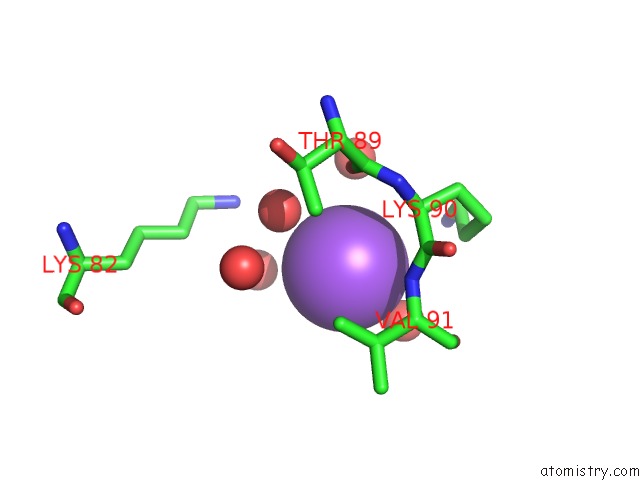
Mono view
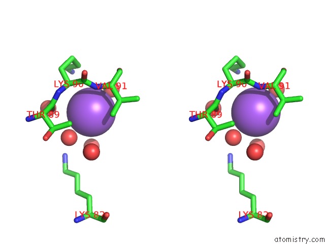
Stereo pair view
Mono view
Stereo pair view
A full contact list of Sodium with other atoms in the Na binding
site number 1 of E. Coli Thioredoxin Variant with PRO76 As Single Proline Residue within 5.0Å range:
|
Reference:
M.Rubini,
M.A.Scharer,
G.Capitani,
R.Glockshuber.
(4R)- and (4S)-Fluoroproline in the Conserved Cis-Prolyl Peptide Bond of the Thioredoxin Fold: Tertiary Structure Context Dictates Ring Puckering. Chembiochem V. 14 1053 2013.
ISSN: ISSN 1439-4227
PubMed: 23712956
DOI: 10.1002/CBIC.201300178
Page generated: Mon Oct 7 15:54:10 2024
ISSN: ISSN 1439-4227
PubMed: 23712956
DOI: 10.1002/CBIC.201300178
Last articles
Ca in 5NERCa in 5NEM
Ca in 5NE5
Ca in 5NBP
Ca in 5NBN
Ca in 5NBM
Ca in 5NBL
Ca in 5N7G
Ca in 5N7F
Ca in 5N7D