Sodium »
PDB 4gxj-4hfc »
4ha4 »
Sodium in PDB 4ha4: Structure of Beta-Glycosidase From Acidilobus Saccharovorans in Complex with Glycerol
Enzymatic activity of Structure of Beta-Glycosidase From Acidilobus Saccharovorans in Complex with Glycerol
All present enzymatic activity of Structure of Beta-Glycosidase From Acidilobus Saccharovorans in Complex with Glycerol:
3.2.1.23;
3.2.1.23;
Protein crystallography data
The structure of Structure of Beta-Glycosidase From Acidilobus Saccharovorans in Complex with Glycerol, PDB code: 4ha4
was solved by
A.A.Trofimov,
K.M.Polyakov,
A.V.Tikhonov,
E.Y.Bezsudnova,
P.V.Dorovatovskii,
V.M.Gumerov,
N.V.Ravin,
K.G.Skryabin,
V.O.Popov,
with X-Ray Crystallography technique. A brief refinement statistics is given in the table below:
| Resolution Low / High (Å) | 19.83 / 1.37 |
| Space group | P 42 21 2 |
| Cell size a, b, c (Å), α, β, γ (°) | 84.120, 84.120, 166.270, 90.00, 90.00, 90.00 |
| R / Rfree (%) | 13 / 16.3 |
Sodium Binding Sites:
The binding sites of Sodium atom in the Structure of Beta-Glycosidase From Acidilobus Saccharovorans in Complex with Glycerol
(pdb code 4ha4). This binding sites where shown within
5.0 Angstroms radius around Sodium atom.
In total only one binding site of Sodium was determined in the Structure of Beta-Glycosidase From Acidilobus Saccharovorans in Complex with Glycerol, PDB code: 4ha4:
In total only one binding site of Sodium was determined in the Structure of Beta-Glycosidase From Acidilobus Saccharovorans in Complex with Glycerol, PDB code: 4ha4:
Sodium binding site 1 out of 1 in 4ha4
Go back to
Sodium binding site 1 out
of 1 in the Structure of Beta-Glycosidase From Acidilobus Saccharovorans in Complex with Glycerol
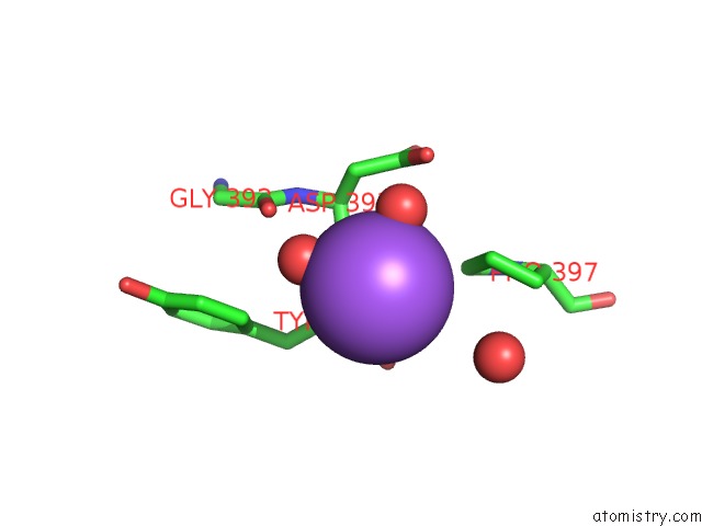
Mono view
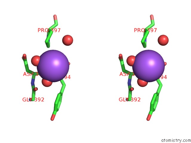
Stereo pair view
Mono view
Stereo pair view
A full contact list of Sodium with other atoms in the Na binding
site number 1 of Structure of Beta-Glycosidase From Acidilobus Saccharovorans in Complex with Glycerol within 5.0Å range:
|
Reference:
A.A.Trofimov,
K.M.Polyakov,
A.V.Tikhonov,
E.Y.Bezsudnova,
P.V.Dorovatovskii,
V.M.Gumerov,
N.V.Ravin,
K.G.Skryabin,
V.O.Popov.
Structures of Beta-Glycosidase From Acidilobus Saccharovorans in Complexes with Tris and Glycerol. Dokl.Biochem.Biophys. V. 449 99.
ISSN: ISSN 1607-6729
PubMed: 23657657
DOI: 10.1134/S1607672913020129
Page generated: Mon Oct 7 15:45:26 2024
ISSN: ISSN 1607-6729
PubMed: 23657657
DOI: 10.1134/S1607672913020129
Last articles
Zn in 9J0NZn in 9J0O
Zn in 9J0P
Zn in 9FJX
Zn in 9EKB
Zn in 9C0F
Zn in 9CAH
Zn in 9CH0
Zn in 9CH3
Zn in 9CH1