Sodium »
PDB 4f5r-4foi »
4fe3 »
Sodium in PDB 4fe3: Structure of Murine Cytosolic 5'-Nucleotidase III Complexed with Uridinine Monophosphate
Enzymatic activity of Structure of Murine Cytosolic 5'-Nucleotidase III Complexed with Uridinine Monophosphate
All present enzymatic activity of Structure of Murine Cytosolic 5'-Nucleotidase III Complexed with Uridinine Monophosphate:
3.1.3.5;
3.1.3.5;
Protein crystallography data
The structure of Structure of Murine Cytosolic 5'-Nucleotidase III Complexed with Uridinine Monophosphate, PDB code: 4fe3
was solved by
E.Bitto,
C.A.Bingman,
with X-Ray Crystallography technique. A brief refinement statistics is given in the table below:
| Resolution Low / High (Å) | 50.00 / 1.74 |
| Space group | P 43 2 2 |
| Cell size a, b, c (Å), α, β, γ (°) | 46.878, 46.878, 287.101, 90.00, 90.00, 90.00 |
| R / Rfree (%) | 16.9 / 21.8 |
Other elements in 4fe3:
The structure of Structure of Murine Cytosolic 5'-Nucleotidase III Complexed with Uridinine Monophosphate also contains other interesting chemical elements:
| Magnesium | (Mg) | 1 atom |
Sodium Binding Sites:
The binding sites of Sodium atom in the Structure of Murine Cytosolic 5'-Nucleotidase III Complexed with Uridinine Monophosphate
(pdb code 4fe3). This binding sites where shown within
5.0 Angstroms radius around Sodium atom.
In total only one binding site of Sodium was determined in the Structure of Murine Cytosolic 5'-Nucleotidase III Complexed with Uridinine Monophosphate, PDB code: 4fe3:
In total only one binding site of Sodium was determined in the Structure of Murine Cytosolic 5'-Nucleotidase III Complexed with Uridinine Monophosphate, PDB code: 4fe3:
Sodium binding site 1 out of 1 in 4fe3
Go back to
Sodium binding site 1 out
of 1 in the Structure of Murine Cytosolic 5'-Nucleotidase III Complexed with Uridinine Monophosphate
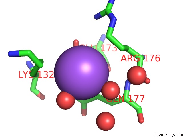
Mono view
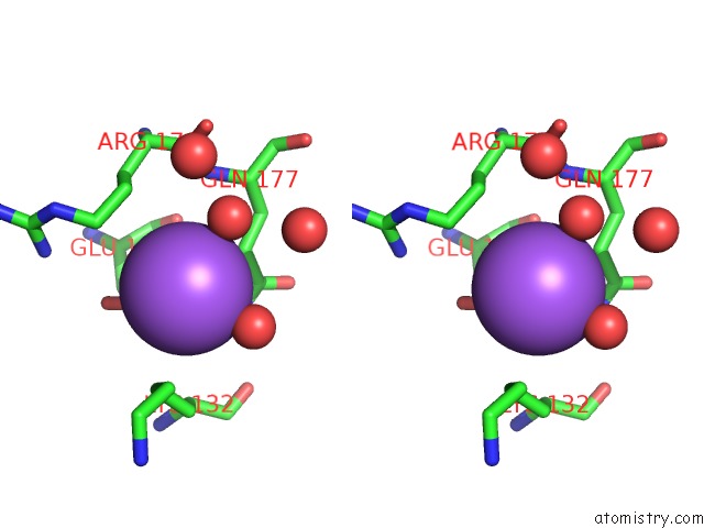
Stereo pair view
Mono view
Stereo pair view
A full contact list of Sodium with other atoms in the Na binding
site number 1 of Structure of Murine Cytosolic 5'-Nucleotidase III Complexed with Uridinine Monophosphate within 5.0Å range:
|
Reference:
C.L.Grobosky,
J.B.Lopez,
S.Rennie,
D.J.Skopelitis,
A.T.Wiest,
C.A.Bingman,
E.Bitto.
Structural Basis of Substrate Specificity and Selectivity of Murine Cytosolic 5'-Nucleotidase III. J.Mol.Biol. V. 423 540 2012.
ISSN: ISSN 0022-2836
PubMed: 22925580
DOI: 10.1016/J.JMB.2012.08.014
Page generated: Mon Oct 7 15:20:14 2024
ISSN: ISSN 0022-2836
PubMed: 22925580
DOI: 10.1016/J.JMB.2012.08.014
Last articles
Zn in 9JYWZn in 9IR4
Zn in 9IR3
Zn in 9GMX
Zn in 9GMW
Zn in 9JEJ
Zn in 9ERF
Zn in 9ERE
Zn in 9EGV
Zn in 9EGW