Sodium »
PDB 4f5r-4foi »
4fax »
Sodium in PDB 4fax: Structure of Oceanobacillus Iheyensis Group II Intron in A Ligand-Free State in the Presence of Na+ and MG2+
Protein crystallography data
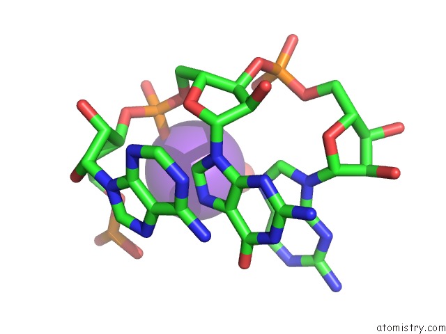
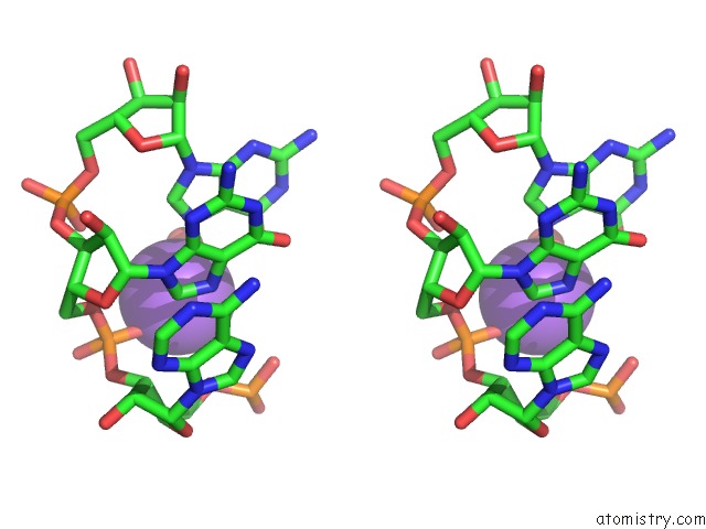
The structure of Structure of Oceanobacillus Iheyensis Group II Intron in A Ligand-Free State in the Presence of Na+ and MG2+, PDB code: 4fax
was solved by
M.Marcia,
A.M.Pyle,
with X-Ray Crystallography technique. A brief refinement statistics is given in the table below:
| Resolution Low / High (Å) | 48.41 / 3.10 |
| Space group | P 21 21 21 |
| Cell size a, b, c (Å), α, β, γ (°) | 89.370, 95.370, 224.750, 90.00, 90.00, 90.00 |
| R / Rfree (%) | 19.6 / 23.8 |
Other elements in 4fax:
The structure of Structure of Oceanobacillus Iheyensis Group II Intron in A Ligand-Free State in the Presence of Na+ and MG2+ also contains other interesting chemical elements:
| Magnesium | (Mg) | 21 atoms |
Sodium Binding Sites:
The binding sites of Sodium atom in the Structure of Oceanobacillus Iheyensis Group II Intron in A Ligand-Free State in the Presence of Na+ and MG2+
(pdb code 4fax). This binding sites where shown within
5.0 Angstroms radius around Sodium atom.
In total 8 binding sites of Sodium where determined in the Structure of Oceanobacillus Iheyensis Group II Intron in A Ligand-Free State in the Presence of Na+ and MG2+, PDB code: 4fax:
Jump to Sodium binding site number: 1; 2; 3; 4; 5; 6; 7; 8;
In total 8 binding sites of Sodium where determined in the Structure of Oceanobacillus Iheyensis Group II Intron in A Ligand-Free State in the Presence of Na+ and MG2+, PDB code: 4fax:
Jump to Sodium binding site number: 1; 2; 3; 4; 5; 6; 7; 8;
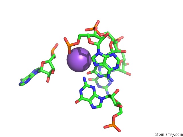
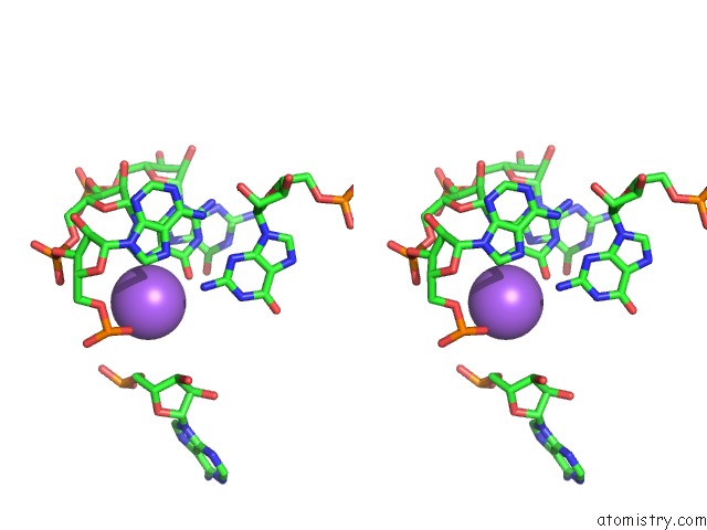
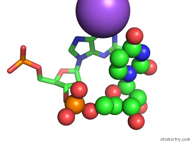
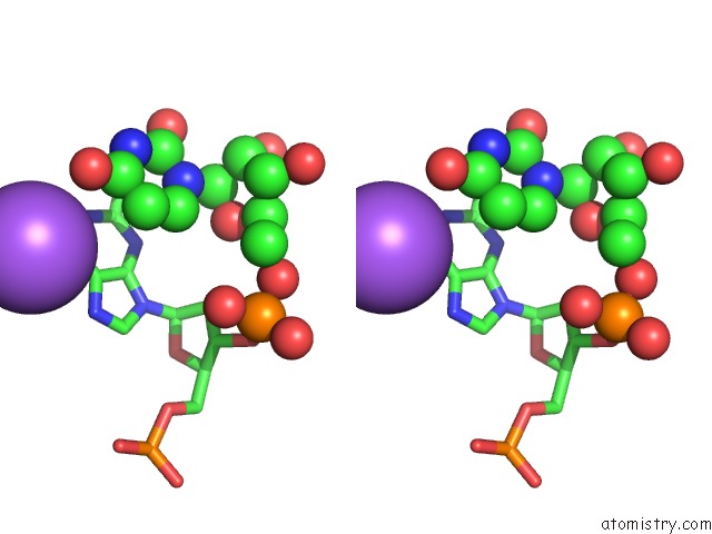
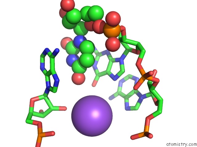
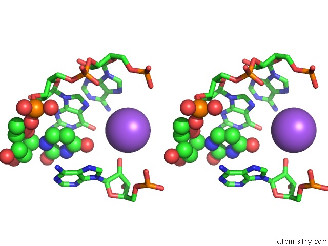
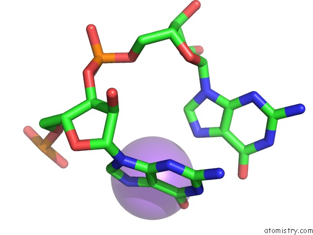
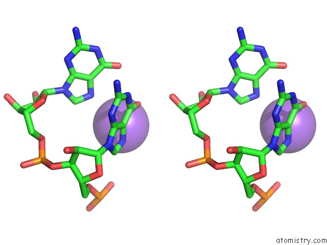
Sodium binding site 1 out of 8 in 4fax
Go back to
Sodium binding site 1 out
of 8 in the Structure of Oceanobacillus Iheyensis Group II Intron in A Ligand-Free State in the Presence of Na+ and MG2+
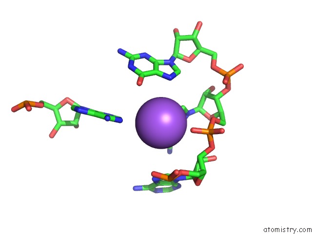
Mono view
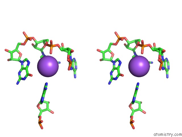
Stereo pair view
Mono view
Stereo pair view
A full contact list of Sodium with other atoms in the Na binding
site number 1 of Structure of Oceanobacillus Iheyensis Group II Intron in A Ligand-Free State in the Presence of Na+ and MG2+ within 5.0Å range:
|
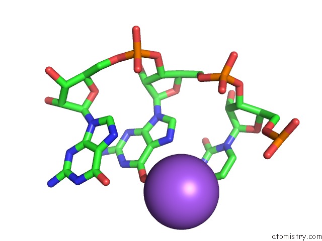
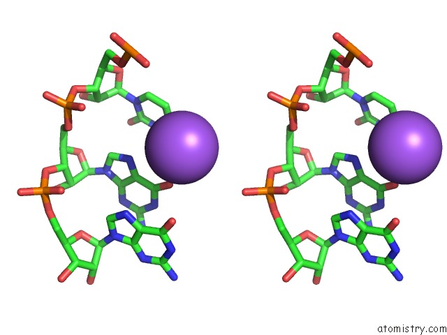
Sodium binding site 2 out of 8 in 4fax
Go back to
Sodium binding site 2 out
of 8 in the Structure of Oceanobacillus Iheyensis Group II Intron in A Ligand-Free State in the Presence of Na+ and MG2+
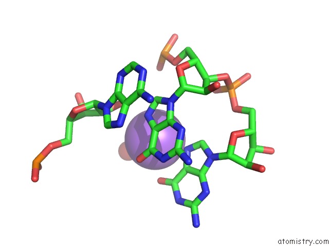
Mono view
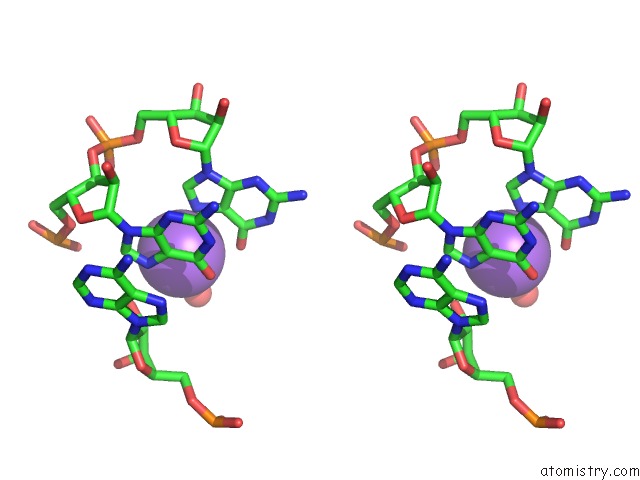
Stereo pair view
Mono view
Stereo pair view
A full contact list of Sodium with other atoms in the Na binding
site number 2 of Structure of Oceanobacillus Iheyensis Group II Intron in A Ligand-Free State in the Presence of Na+ and MG2+ within 5.0Å range:
|
Sodium binding site 3 out of 8 in 4fax
Go back to
Sodium binding site 3 out
of 8 in the Structure of Oceanobacillus Iheyensis Group II Intron in A Ligand-Free State in the Presence of Na+ and MG2+
Mono view
Stereo pair view
Mono view
Stereo pair view
A full contact list of Sodium with other atoms in the Na binding
site number 3 of Structure of Oceanobacillus Iheyensis Group II Intron in A Ligand-Free State in the Presence of Na+ and MG2+ within 5.0Å range:
|
Sodium binding site 4 out of 8 in 4fax
Go back to
Sodium binding site 4 out
of 8 in the Structure of Oceanobacillus Iheyensis Group II Intron in A Ligand-Free State in the Presence of Na+ and MG2+
Mono view
Stereo pair view
Mono view
Stereo pair view
A full contact list of Sodium with other atoms in the Na binding
site number 4 of Structure of Oceanobacillus Iheyensis Group II Intron in A Ligand-Free State in the Presence of Na+ and MG2+ within 5.0Å range:
|
Sodium binding site 5 out of 8 in 4fax
Go back to
Sodium binding site 5 out
of 8 in the Structure of Oceanobacillus Iheyensis Group II Intron in A Ligand-Free State in the Presence of Na+ and MG2+
Mono view
Stereo pair view
Mono view
Stereo pair view
A full contact list of Sodium with other atoms in the Na binding
site number 5 of Structure of Oceanobacillus Iheyensis Group II Intron in A Ligand-Free State in the Presence of Na+ and MG2+ within 5.0Å range:
|
Sodium binding site 6 out of 8 in 4fax
Go back to
Sodium binding site 6 out
of 8 in the Structure of Oceanobacillus Iheyensis Group II Intron in A Ligand-Free State in the Presence of Na+ and MG2+
Mono view
Stereo pair view
Mono view
Stereo pair view
A full contact list of Sodium with other atoms in the Na binding
site number 6 of Structure of Oceanobacillus Iheyensis Group II Intron in A Ligand-Free State in the Presence of Na+ and MG2+ within 5.0Å range:
|
Sodium binding site 7 out of 8 in 4fax
Go back to
Sodium binding site 7 out
of 8 in the Structure of Oceanobacillus Iheyensis Group II Intron in A Ligand-Free State in the Presence of Na+ and MG2+
Mono view
Stereo pair view
Mono view
Stereo pair view
A full contact list of Sodium with other atoms in the Na binding
site number 7 of Structure of Oceanobacillus Iheyensis Group II Intron in A Ligand-Free State in the Presence of Na+ and MG2+ within 5.0Å range:
|
Sodium binding site 8 out of 8 in 4fax
Go back to
Sodium binding site 8 out
of 8 in the Structure of Oceanobacillus Iheyensis Group II Intron in A Ligand-Free State in the Presence of Na+ and MG2+
Mono view
Stereo pair view
Mono view
Stereo pair view
A full contact list of Sodium with other atoms in the Na binding
site number 8 of Structure of Oceanobacillus Iheyensis Group II Intron in A Ligand-Free State in the Presence of Na+ and MG2+ within 5.0Å range:
|
Reference:
M.Marcia,
A.M.Pyle.
Visualizing Group II Intron Catalysis Through the Stages of Splicing. Cell(Cambridge,Mass.) V. 151 497 2012.
ISSN: ISSN 0092-8674
PubMed: 23101623
DOI: 10.1016/J.CELL.2012.09.033
Page generated: Mon Oct 7 15:20:05 2024
ISSN: ISSN 0092-8674
PubMed: 23101623
DOI: 10.1016/J.CELL.2012.09.033
Last articles
Zn in 9MJ5Zn in 9HNW
Zn in 9G0L
Zn in 9FNE
Zn in 9DZN
Zn in 9E0I
Zn in 9D32
Zn in 9DAK
Zn in 8ZXC
Zn in 8ZUF