Sodium »
PDB 4ehu-4f5q »
4ekf »
Sodium in PDB 4ekf: Structure of the Inactive Adenovirus Proteinase at 0.98 Angstrom Resolution
Enzymatic activity of Structure of the Inactive Adenovirus Proteinase at 0.98 Angstrom Resolution
All present enzymatic activity of Structure of the Inactive Adenovirus Proteinase at 0.98 Angstrom Resolution:
3.4.22.39;
3.4.22.39;
Protein crystallography data
The structure of Structure of the Inactive Adenovirus Proteinase at 0.98 Angstrom Resolution, PDB code: 4ekf
was solved by
M.L.Baniecki,
W.J.Mcgrath,
W.F.Mangel,
with X-Ray Crystallography technique. A brief refinement statistics is given in the table below:
| Resolution Low / High (Å) | 20.00 / 0.98 |
| Space group | P 1 21 1 |
| Cell size a, b, c (Å), α, β, γ (°) | 36.270, 54.540, 42.410, 90.00, 100.10, 90.00 |
| R / Rfree (%) | 13.5 / 16.8 |
Sodium Binding Sites:
The binding sites of Sodium atom in the Structure of the Inactive Adenovirus Proteinase at 0.98 Angstrom Resolution
(pdb code 4ekf). This binding sites where shown within
5.0 Angstroms radius around Sodium atom.
In total only one binding site of Sodium was determined in the Structure of the Inactive Adenovirus Proteinase at 0.98 Angstrom Resolution, PDB code: 4ekf:
In total only one binding site of Sodium was determined in the Structure of the Inactive Adenovirus Proteinase at 0.98 Angstrom Resolution, PDB code: 4ekf:
Sodium binding site 1 out of 1 in 4ekf
Go back to
Sodium binding site 1 out
of 1 in the Structure of the Inactive Adenovirus Proteinase at 0.98 Angstrom Resolution
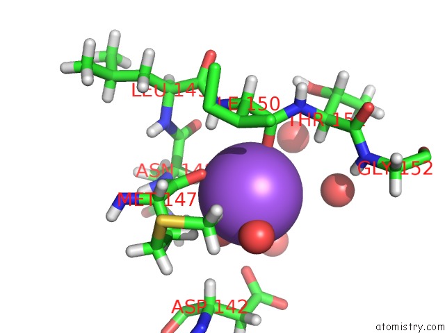
Mono view
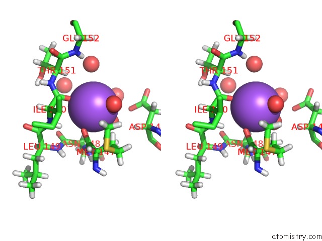
Stereo pair view
Mono view
Stereo pair view
A full contact list of Sodium with other atoms in the Na binding
site number 1 of Structure of the Inactive Adenovirus Proteinase at 0.98 Angstrom Resolution within 5.0Å range:
|
Reference:
M.L.Baniecki,
W.J.Mcgrath,
W.F.Mangel.
Regulation of A Viral Proteinase By A Peptide and Dna in One-Dimensional Space: III. Atomic Resolution Structure of the Nascent Form of the Adenovirus Proteinase. J.Biol.Chem. V. 288 2081 2013.
ISSN: ISSN 0021-9258
PubMed: 23043139
DOI: 10.1074/JBC.M112.407429
Page generated: Mon Oct 7 15:10:20 2024
ISSN: ISSN 0021-9258
PubMed: 23043139
DOI: 10.1074/JBC.M112.407429
Last articles
Zn in 9J0NZn in 9J0O
Zn in 9J0P
Zn in 9FJX
Zn in 9EKB
Zn in 9C0F
Zn in 9CAH
Zn in 9CH0
Zn in 9CH3
Zn in 9CH1