Sodium »
PDB 4coo-4ddo »
4cuo »
Sodium in PDB 4cuo: Banyan Peroxidase with Glycosylation
Enzymatic activity of Banyan Peroxidase with Glycosylation
All present enzymatic activity of Banyan Peroxidase with Glycosylation:
1.11.1.7;
1.11.1.7;
Protein crystallography data
The structure of Banyan Peroxidase with Glycosylation, PDB code: 4cuo
was solved by
G.J.Palm,
A.Sharma,
W.Hinrichs,
with X-Ray Crystallography technique. A brief refinement statistics is given in the table below:
| Resolution Low / High (Å) | 63.32 / 1.67 |
| Space group | P 32 2 1 |
| Cell size a, b, c (Å), α, β, γ (°) | 73.115, 73.115, 164.596, 90.00, 90.00, 120.00 |
| R / Rfree (%) | 15.654 / 18.029 |
Other elements in 4cuo:
The structure of Banyan Peroxidase with Glycosylation also contains other interesting chemical elements:
| Iron | (Fe) | 1 atom |
| Chlorine | (Cl) | 3 atoms |
| Calcium | (Ca) | 2 atoms |
Sodium Binding Sites:
The binding sites of Sodium atom in the Banyan Peroxidase with Glycosylation
(pdb code 4cuo). This binding sites where shown within
5.0 Angstroms radius around Sodium atom.
In total only one binding site of Sodium was determined in the Banyan Peroxidase with Glycosylation, PDB code: 4cuo:
In total only one binding site of Sodium was determined in the Banyan Peroxidase with Glycosylation, PDB code: 4cuo:
Sodium binding site 1 out of 1 in 4cuo
Go back to
Sodium binding site 1 out
of 1 in the Banyan Peroxidase with Glycosylation
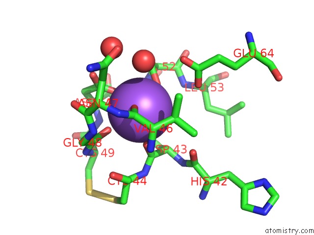
Mono view
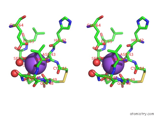
Stereo pair view
Mono view
Stereo pair view
A full contact list of Sodium with other atoms in the Na binding
site number 1 of Banyan Peroxidase with Glycosylation within 5.0Å range:
|
Reference:
G.J.Palm,
A.Sharma,
M.Kumari,
S.Panjikar,
D.Albrecht,
M.V.Jagannadham,
W.Hinrichs.
Post-Translational Modification and Extended Glycosylation Pattern of A Plant Latex Peroxidase of Native Source Characterized By X-Ray Crystallography. Febs J. V. 281 4319 2014.
ISSN: ISSN 1742-464X
PubMed: 24980207
DOI: 10.1111/FEBS.12900
Page generated: Mon Oct 7 14:48:12 2024
ISSN: ISSN 1742-464X
PubMed: 24980207
DOI: 10.1111/FEBS.12900
Last articles
Fe in 2YXOFe in 2YRS
Fe in 2YXC
Fe in 2YNM
Fe in 2YVJ
Fe in 2YP1
Fe in 2YU2
Fe in 2YU1
Fe in 2YQB
Fe in 2YOO