Sodium »
PDB 4c6s-4cnt »
4ciw »
Sodium in PDB 4ciw: Crystal Structure of Mycobacterium Tuberculosis Type 2 Dehydroquinase in Complex with (1R,4R,5R)-1,4,5-Trihydroxy-3- (2-Hydroxy)Ethylcyclohex-2-Ene-1-Carboxylic Acid
Enzymatic activity of Crystal Structure of Mycobacterium Tuberculosis Type 2 Dehydroquinase in Complex with (1R,4R,5R)-1,4,5-Trihydroxy-3- (2-Hydroxy)Ethylcyclohex-2-Ene-1-Carboxylic Acid
All present enzymatic activity of Crystal Structure of Mycobacterium Tuberculosis Type 2 Dehydroquinase in Complex with (1R,4R,5R)-1,4,5-Trihydroxy-3- (2-Hydroxy)Ethylcyclohex-2-Ene-1-Carboxylic Acid:
4.2.1.10;
4.2.1.10;
Protein crystallography data
The structure of Crystal Structure of Mycobacterium Tuberculosis Type 2 Dehydroquinase in Complex with (1R,4R,5R)-1,4,5-Trihydroxy-3- (2-Hydroxy)Ethylcyclohex-2-Ene-1-Carboxylic Acid, PDB code: 4ciw
was solved by
J.M.Otero,
A.L.Llamas-Saiz,
H.Lamb,
A.R.Hawkins,
B.Blanco,
A.Sedes,
A.Peon,
C.Gonzalez-Bello,
M.J.Van Raaij,
with X-Ray Crystallography technique. A brief refinement statistics is given in the table below:
| Resolution Low / High (Å) | 38.13 / 2.20 |
| Space group | F 2 3 |
| Cell size a, b, c (Å), α, β, γ (°) | 126.380, 126.380, 126.380, 90.00, 90.00, 90.00 |
| R / Rfree (%) | 14.177 / 19.227 |
Sodium Binding Sites:
The binding sites of Sodium atom in the Crystal Structure of Mycobacterium Tuberculosis Type 2 Dehydroquinase in Complex with (1R,4R,5R)-1,4,5-Trihydroxy-3- (2-Hydroxy)Ethylcyclohex-2-Ene-1-Carboxylic Acid
(pdb code 4ciw). This binding sites where shown within
5.0 Angstroms radius around Sodium atom.
In total 3 binding sites of Sodium where determined in the Crystal Structure of Mycobacterium Tuberculosis Type 2 Dehydroquinase in Complex with (1R,4R,5R)-1,4,5-Trihydroxy-3- (2-Hydroxy)Ethylcyclohex-2-Ene-1-Carboxylic Acid, PDB code: 4ciw:
Jump to Sodium binding site number: 1; 2; 3;
In total 3 binding sites of Sodium where determined in the Crystal Structure of Mycobacterium Tuberculosis Type 2 Dehydroquinase in Complex with (1R,4R,5R)-1,4,5-Trihydroxy-3- (2-Hydroxy)Ethylcyclohex-2-Ene-1-Carboxylic Acid, PDB code: 4ciw:
Jump to Sodium binding site number: 1; 2; 3;
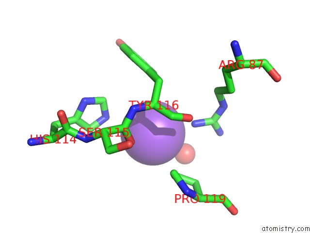
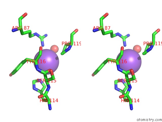
Sodium binding site 1 out of 3 in 4ciw
Go back to
Sodium binding site 1 out
of 3 in the Crystal Structure of Mycobacterium Tuberculosis Type 2 Dehydroquinase in Complex with (1R,4R,5R)-1,4,5-Trihydroxy-3- (2-Hydroxy)Ethylcyclohex-2-Ene-1-Carboxylic Acid
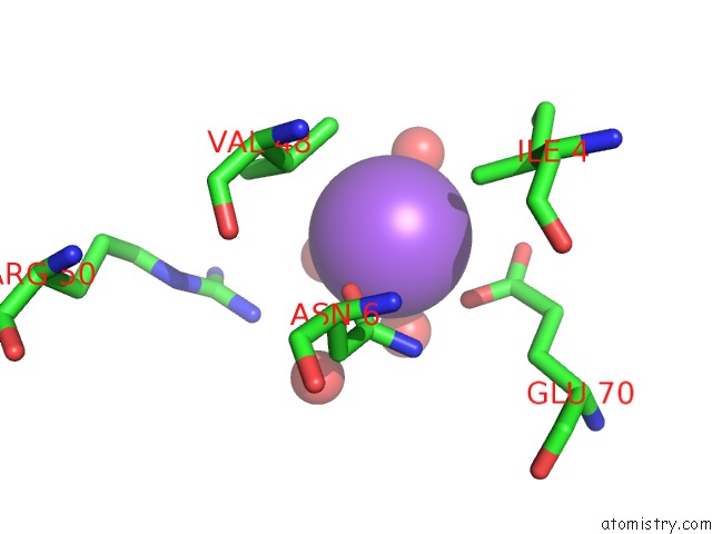
Mono view
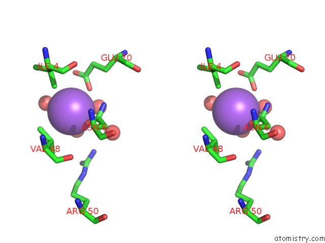
Stereo pair view
Mono view
Stereo pair view
A full contact list of Sodium with other atoms in the Na binding
site number 1 of Crystal Structure of Mycobacterium Tuberculosis Type 2 Dehydroquinase in Complex with (1R,4R,5R)-1,4,5-Trihydroxy-3- (2-Hydroxy)Ethylcyclohex-2-Ene-1-Carboxylic Acid within 5.0Å range:
|
Sodium binding site 2 out of 3 in 4ciw
Go back to
Sodium binding site 2 out
of 3 in the Crystal Structure of Mycobacterium Tuberculosis Type 2 Dehydroquinase in Complex with (1R,4R,5R)-1,4,5-Trihydroxy-3- (2-Hydroxy)Ethylcyclohex-2-Ene-1-Carboxylic Acid
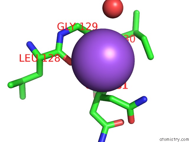
Mono view
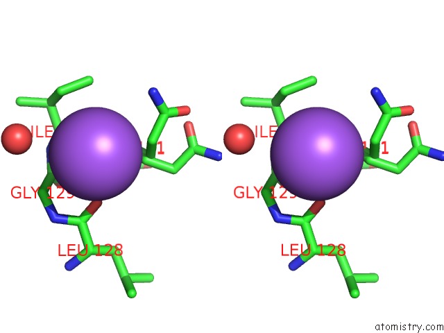
Stereo pair view
Mono view
Stereo pair view
A full contact list of Sodium with other atoms in the Na binding
site number 2 of Crystal Structure of Mycobacterium Tuberculosis Type 2 Dehydroquinase in Complex with (1R,4R,5R)-1,4,5-Trihydroxy-3- (2-Hydroxy)Ethylcyclohex-2-Ene-1-Carboxylic Acid within 5.0Å range:
|
Sodium binding site 3 out of 3 in 4ciw
Go back to
Sodium binding site 3 out
of 3 in the Crystal Structure of Mycobacterium Tuberculosis Type 2 Dehydroquinase in Complex with (1R,4R,5R)-1,4,5-Trihydroxy-3- (2-Hydroxy)Ethylcyclohex-2-Ene-1-Carboxylic Acid
Mono view
Stereo pair view
Mono view
Stereo pair view
A full contact list of Sodium with other atoms in the Na binding
site number 3 of Crystal Structure of Mycobacterium Tuberculosis Type 2 Dehydroquinase in Complex with (1R,4R,5R)-1,4,5-Trihydroxy-3- (2-Hydroxy)Ethylcyclohex-2-Ene-1-Carboxylic Acid within 5.0Å range:
|
Reference:
B.Blanco,
A.Sedes,
A.Peon,
J.M.Otero,
M.J.Van Raaij,
P.Thompson,
A.R.Hawkins,
C.Gonzalez-Bello.
Exploring the Water-Binding Pocket of the Type II Dehydroquinase Enzyme in the Structure-Based Design of Inhibitors. J.Med.Chem. V. 57 3494 2014.
ISSN: ISSN 0022-2623
PubMed: 24689821
DOI: 10.1021/JM500175Z
Page generated: Sun Aug 17 18:48:52 2025
ISSN: ISSN 0022-2623
PubMed: 24689821
DOI: 10.1021/JM500175Z
Last articles
Na in 4OMGNa in 4OLF
Na in 4OLN
Na in 4OHC
Na in 4OGL
Na in 4OJ1
Na in 4OFI
Na in 4OIC
Na in 4OF8
Na in 4OFX