Sodium »
PDB 4c6s-4cnt »
4ch2 »
Sodium in PDB 4ch2: Low-Salt Crystal Structure of A Thrombin-Gpibalpha Peptide Complex
Enzymatic activity of Low-Salt Crystal Structure of A Thrombin-Gpibalpha Peptide Complex
All present enzymatic activity of Low-Salt Crystal Structure of A Thrombin-Gpibalpha Peptide Complex:
3.4.21.5;
3.4.21.5;
Protein crystallography data
The structure of Low-Salt Crystal Structure of A Thrombin-Gpibalpha Peptide Complex, PDB code: 4ch2
was solved by
B.C.Lechtenberg,
S.M.V.Freund,
J.A.Huntington,
with X-Ray Crystallography technique. A brief refinement statistics is given in the table below:
| Resolution Low / High (Å) | 37.10 / 1.60 |
| Space group | C 1 2 1 |
| Cell size a, b, c (Å), α, β, γ (°) | 149.570, 50.560, 76.280, 90.00, 97.15, 90.00 |
| R / Rfree (%) | 15.452 / 18.73 |
Sodium Binding Sites:
The binding sites of Sodium atom in the Low-Salt Crystal Structure of A Thrombin-Gpibalpha Peptide Complex
(pdb code 4ch2). This binding sites where shown within
5.0 Angstroms radius around Sodium atom.
In total 2 binding sites of Sodium where determined in the Low-Salt Crystal Structure of A Thrombin-Gpibalpha Peptide Complex, PDB code: 4ch2:
Jump to Sodium binding site number: 1; 2;
In total 2 binding sites of Sodium where determined in the Low-Salt Crystal Structure of A Thrombin-Gpibalpha Peptide Complex, PDB code: 4ch2:
Jump to Sodium binding site number: 1; 2;
Sodium binding site 1 out of 2 in 4ch2
Go back to
Sodium binding site 1 out
of 2 in the Low-Salt Crystal Structure of A Thrombin-Gpibalpha Peptide Complex
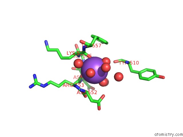
Mono view
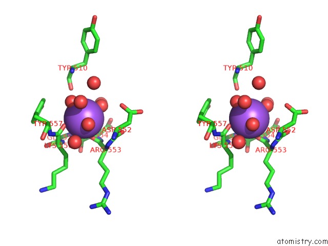
Stereo pair view
Mono view
Stereo pair view
A full contact list of Sodium with other atoms in the Na binding
site number 1 of Low-Salt Crystal Structure of A Thrombin-Gpibalpha Peptide Complex within 5.0Å range:
|
Sodium binding site 2 out of 2 in 4ch2
Go back to
Sodium binding site 2 out
of 2 in the Low-Salt Crystal Structure of A Thrombin-Gpibalpha Peptide Complex
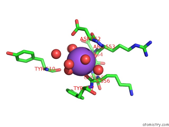
Mono view
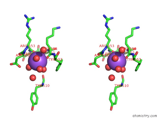
Stereo pair view
Mono view
Stereo pair view
A full contact list of Sodium with other atoms in the Na binding
site number 2 of Low-Salt Crystal Structure of A Thrombin-Gpibalpha Peptide Complex within 5.0Å range:
|
Reference:
B.C.Lechtenberg,
S.M.V.Freund,
J.A.Huntington.
Gpibalpha Interacts Exclusively with Exosite II of Thrombin J.Mol.Biol. V. 426 881 2014.
ISSN: ISSN 0022-2836
PubMed: 24316004
DOI: 10.1016/J.JMB.2013.11.027
Page generated: Mon Oct 7 14:43:41 2024
ISSN: ISSN 0022-2836
PubMed: 24316004
DOI: 10.1016/J.JMB.2013.11.027
Last articles
Zn in 9MJ5Zn in 9HNW
Zn in 9G0L
Zn in 9FNE
Zn in 9DZN
Zn in 9E0I
Zn in 9D32
Zn in 9DAK
Zn in 8ZXC
Zn in 8ZUF