Sodium »
PDB 3zux-4adj »
4adi »
Sodium in PDB 4adi: Crystal Structure of the Rubella Virus Envelope Glycoprotein E1 in Post-Fusion Form (Crystal Form I)
Protein crystallography data
The structure of Crystal Structure of the Rubella Virus Envelope Glycoprotein E1 in Post-Fusion Form (Crystal Form I), PDB code: 4adi
was solved by
R.M.Dubois,
M.C.Vaney,
M.A.Tortorici,
R.Al Kurdi,
G.Barba-Spaeth,
F.A.Rey,
with X-Ray Crystallography technique. A brief refinement statistics is given in the table below:
| Resolution Low / High (Å) | 20.00 / 1.80 |
| Space group | P 21 21 21 |
| Cell size a, b, c (Å), α, β, γ (°) | 129.980, 121.380, 126.430, 90.00, 90.00, 90.00 |
| R / Rfree (%) | 16.66 / 18.31 |
Sodium Binding Sites:
The binding sites of Sodium atom in the Crystal Structure of the Rubella Virus Envelope Glycoprotein E1 in Post-Fusion Form (Crystal Form I)
(pdb code 4adi). This binding sites where shown within
5.0 Angstroms radius around Sodium atom.
In total 3 binding sites of Sodium where determined in the Crystal Structure of the Rubella Virus Envelope Glycoprotein E1 in Post-Fusion Form (Crystal Form I), PDB code: 4adi:
Jump to Sodium binding site number: 1; 2; 3;
In total 3 binding sites of Sodium where determined in the Crystal Structure of the Rubella Virus Envelope Glycoprotein E1 in Post-Fusion Form (Crystal Form I), PDB code: 4adi:
Jump to Sodium binding site number: 1; 2; 3;
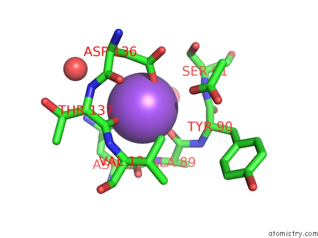
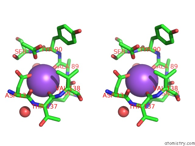
Sodium binding site 1 out of 3 in 4adi
Go back to
Sodium binding site 1 out
of 3 in the Crystal Structure of the Rubella Virus Envelope Glycoprotein E1 in Post-Fusion Form (Crystal Form I)
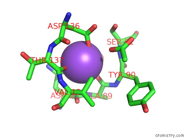
Mono view
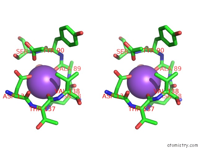
Stereo pair view
Mono view
Stereo pair view
A full contact list of Sodium with other atoms in the Na binding
site number 1 of Crystal Structure of the Rubella Virus Envelope Glycoprotein E1 in Post-Fusion Form (Crystal Form I) within 5.0Å range:
|
Sodium binding site 2 out of 3 in 4adi
Go back to
Sodium binding site 2 out
of 3 in the Crystal Structure of the Rubella Virus Envelope Glycoprotein E1 in Post-Fusion Form (Crystal Form I)
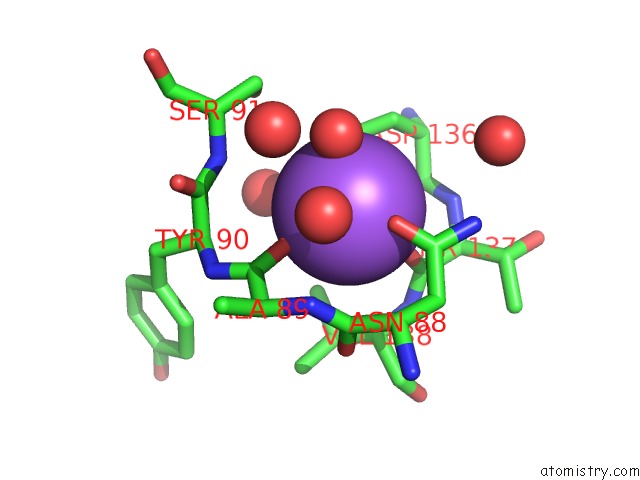
Mono view
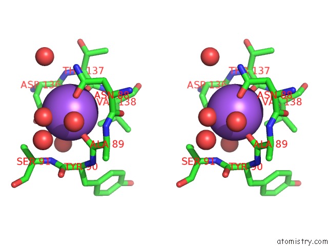
Stereo pair view
Mono view
Stereo pair view
A full contact list of Sodium with other atoms in the Na binding
site number 2 of Crystal Structure of the Rubella Virus Envelope Glycoprotein E1 in Post-Fusion Form (Crystal Form I) within 5.0Å range:
|
Sodium binding site 3 out of 3 in 4adi
Go back to
Sodium binding site 3 out
of 3 in the Crystal Structure of the Rubella Virus Envelope Glycoprotein E1 in Post-Fusion Form (Crystal Form I)
Mono view
Stereo pair view
Mono view
Stereo pair view
A full contact list of Sodium with other atoms in the Na binding
site number 3 of Crystal Structure of the Rubella Virus Envelope Glycoprotein E1 in Post-Fusion Form (Crystal Form I) within 5.0Å range:
|
Reference:
R.M.Dubois,
M.C.Vaney,
M.A.Tortorici,
R.A.Kurdi,
G.Barba-Spaeth,
T.Krey,
F.A.Rey.
Functional and Evolutionary Insight From the Crystal Structure of Rubella Virus Protein E1. Nature V. 493 552 2013.
ISSN: ISSN 0028-0836
PubMed: 23292515
DOI: 10.1038/NATURE11741
Page generated: Mon Oct 7 14:19:54 2024
ISSN: ISSN 0028-0836
PubMed: 23292515
DOI: 10.1038/NATURE11741
Last articles
Zn in 9MJ5Zn in 9HNW
Zn in 9G0L
Zn in 9FNE
Zn in 9DZN
Zn in 9E0I
Zn in 9D32
Zn in 9DAK
Zn in 8ZXC
Zn in 8ZUF