Sodium »
PDB 3zux-4adj »
4a6u »
Sodium in PDB 4a6u: Crystal Structure of the Omega Transaminase From Chromobacterium Violaceum in the Apo Form, Crystallised From Peg 3350
Enzymatic activity of Crystal Structure of the Omega Transaminase From Chromobacterium Violaceum in the Apo Form, Crystallised From Peg 3350
All present enzymatic activity of Crystal Structure of the Omega Transaminase From Chromobacterium Violaceum in the Apo Form, Crystallised From Peg 3350:
2.6.1.18; 2.6.1.62;
2.6.1.18; 2.6.1.62;
Protein crystallography data
The structure of Crystal Structure of the Omega Transaminase From Chromobacterium Violaceum in the Apo Form, Crystallised From Peg 3350, PDB code: 4a6u
was solved by
D.T.Logan,
M.Hakansson,
K.Yengo,
M.Svedendahl Humble,
K.Engelmarkcassimjee,
B.Walse,
V.Abedi,
H.-J.Federsel,
P.Berglund,
with X-Ray Crystallography technique. A brief refinement statistics is given in the table below:
| Resolution Low / High (Å) | 29.47 / 1.69 |
| Space group | P 1 |
| Cell size a, b, c (Å), α, β, γ (°) | 57.170, 61.110, 60.240, 109.14, 85.59, 103.25 |
| R / Rfree (%) | 15.5 / 18 |
Sodium Binding Sites:
The binding sites of Sodium atom in the Crystal Structure of the Omega Transaminase From Chromobacterium Violaceum in the Apo Form, Crystallised From Peg 3350
(pdb code 4a6u). This binding sites where shown within
5.0 Angstroms radius around Sodium atom.
In total 2 binding sites of Sodium where determined in the Crystal Structure of the Omega Transaminase From Chromobacterium Violaceum in the Apo Form, Crystallised From Peg 3350, PDB code: 4a6u:
Jump to Sodium binding site number: 1; 2;
In total 2 binding sites of Sodium where determined in the Crystal Structure of the Omega Transaminase From Chromobacterium Violaceum in the Apo Form, Crystallised From Peg 3350, PDB code: 4a6u:
Jump to Sodium binding site number: 1; 2;
Sodium binding site 1 out of 2 in 4a6u
Go back to
Sodium binding site 1 out
of 2 in the Crystal Structure of the Omega Transaminase From Chromobacterium Violaceum in the Apo Form, Crystallised From Peg 3350
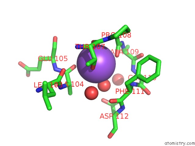
Mono view
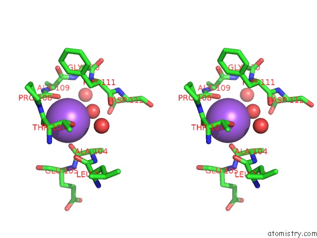
Stereo pair view
Mono view
Stereo pair view
A full contact list of Sodium with other atoms in the Na binding
site number 1 of Crystal Structure of the Omega Transaminase From Chromobacterium Violaceum in the Apo Form, Crystallised From Peg 3350 within 5.0Å range:
|
Sodium binding site 2 out of 2 in 4a6u
Go back to
Sodium binding site 2 out
of 2 in the Crystal Structure of the Omega Transaminase From Chromobacterium Violaceum in the Apo Form, Crystallised From Peg 3350
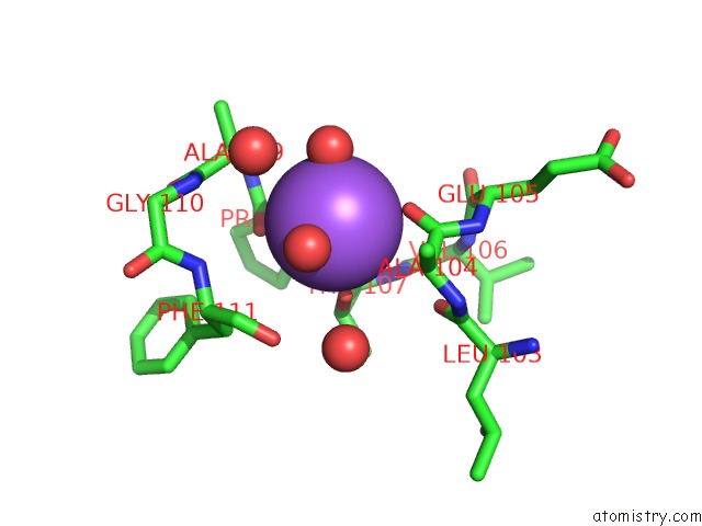
Mono view
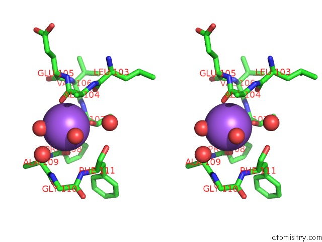
Stereo pair view
Mono view
Stereo pair view
A full contact list of Sodium with other atoms in the Na binding
site number 2 of Crystal Structure of the Omega Transaminase From Chromobacterium Violaceum in the Apo Form, Crystallised From Peg 3350 within 5.0Å range:
|
Reference:
M.Svedendahl Humble,
K.Engelmark Cassimjee,
M.Hakansson,
Y.R.Kimbung,
B.Walse,
V.Abedi,
H.-J.Federsel,
P.Berglund,
D.T.Logan.
Crystal Structures of the Chromobacterium Violaceum Omega-Transaminase Reveal Major Structural Rearrangements Upon Binding of Coenzyme Plp. Febs J. V. 279 779 2012.
ISSN: ISSN 1742-464X
PubMed: 22268978
DOI: 10.1111/J.1742-4658.2011.08468.X
Page generated: Mon Oct 7 14:18:37 2024
ISSN: ISSN 1742-464X
PubMed: 22268978
DOI: 10.1111/J.1742-4658.2011.08468.X
Last articles
Zn in 9MJ5Zn in 9HNW
Zn in 9G0L
Zn in 9FNE
Zn in 9DZN
Zn in 9E0I
Zn in 9D32
Zn in 9DAK
Zn in 8ZXC
Zn in 8ZUF