Sodium »
PDB 3zux-4adj »
435d »
Sodium in PDB 435d: 5'-R(*Up*Ap*Gp*Cp*Cp*Cp*C)-3', 5'-R(*Gp*Gp*Gp*Gp*Cp*Up*A)-3'
Protein crystallography data
The structure of 5'-R(*Up*Ap*Gp*Cp*Cp*Cp*C)-3', 5'-R(*Gp*Gp*Gp*Gp*Cp*Up*A)-3', PDB code: 435d
was solved by
U.Mueller,
H.Schuebel,
M.Sprinzl,
U.Heinemann,
with X-Ray Crystallography technique. A brief refinement statistics is given in the table below:
| Resolution Low / High (Å) | 15.00 / 1.40 |
| Space group | P 1 |
| Cell size a, b, c (Å), α, β, γ (°) | 26.680, 26.697, 30.459, 104.29, 104.22, 91.66 |
| R / Rfree (%) | 15.8 / 21.7 |
Sodium Binding Sites:
The binding sites of Sodium atom in the 5'-R(*Up*Ap*Gp*Cp*Cp*Cp*C)-3', 5'-R(*Gp*Gp*Gp*Gp*Cp*Up*A)-3'
(pdb code 435d). This binding sites where shown within
5.0 Angstroms radius around Sodium atom.
In total 2 binding sites of Sodium where determined in the 5'-R(*Up*Ap*Gp*Cp*Cp*Cp*C)-3', 5'-R(*Gp*Gp*Gp*Gp*Cp*Up*A)-3', PDB code: 435d:
Jump to Sodium binding site number: 1; 2;
In total 2 binding sites of Sodium where determined in the 5'-R(*Up*Ap*Gp*Cp*Cp*Cp*C)-3', 5'-R(*Gp*Gp*Gp*Gp*Cp*Up*A)-3', PDB code: 435d:
Jump to Sodium binding site number: 1; 2;
Sodium binding site 1 out of 2 in 435d
Go back to
Sodium binding site 1 out
of 2 in the 5'-R(*Up*Ap*Gp*Cp*Cp*Cp*C)-3', 5'-R(*Gp*Gp*Gp*Gp*Cp*Up*A)-3'
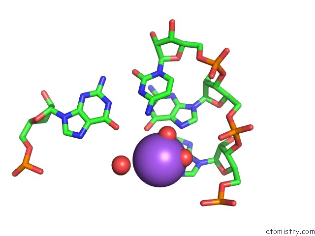
Mono view
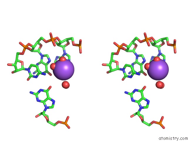
Stereo pair view
Mono view
Stereo pair view
A full contact list of Sodium with other atoms in the Na binding
site number 1 of 5'-R(*Up*Ap*Gp*Cp*Cp*Cp*C)-3', 5'-R(*Gp*Gp*Gp*Gp*Cp*Up*A)-3' within 5.0Å range:
|
Sodium binding site 2 out of 2 in 435d
Go back to
Sodium binding site 2 out
of 2 in the 5'-R(*Up*Ap*Gp*Cp*Cp*Cp*C)-3', 5'-R(*Gp*Gp*Gp*Gp*Cp*Up*A)-3'
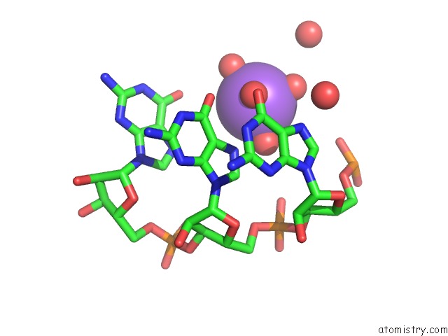
Mono view
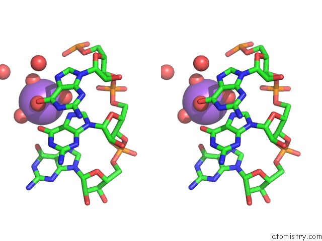
Stereo pair view
Mono view
Stereo pair view
A full contact list of Sodium with other atoms in the Na binding
site number 2 of 5'-R(*Up*Ap*Gp*Cp*Cp*Cp*C)-3', 5'-R(*Gp*Gp*Gp*Gp*Cp*Up*A)-3' within 5.0Å range:
|
Reference:
U.Mueller,
H.Schubel,
M.Sprinzl,
U.Heinemann.
Crystal Structure of Acceptor Stem of Trna(Ala) From Escherichia Coli Shows Unique G.U Wobble Base Pair at 1.16 A Resolution. Rna V. 5 670 1999.
ISSN: ISSN 1355-8382
PubMed: 10334337
DOI: 10.1017/S1355838299982304
Page generated: Mon Oct 7 14:16:09 2024
ISSN: ISSN 1355-8382
PubMed: 10334337
DOI: 10.1017/S1355838299982304
Last articles
Zn in 9MJ5Zn in 9HNW
Zn in 9G0L
Zn in 9FNE
Zn in 9DZN
Zn in 9E0I
Zn in 9D32
Zn in 9DAK
Zn in 8ZXC
Zn in 8ZUF