Sodium »
PDB 3zux-4adj »
3zyv »
Sodium in PDB 3zyv: Crystal Structure of the Mouse Liver Aldehyde Oxidase 3 (MAOX3)
Enzymatic activity of Crystal Structure of the Mouse Liver Aldehyde Oxidase 3 (MAOX3)
All present enzymatic activity of Crystal Structure of the Mouse Liver Aldehyde Oxidase 3 (MAOX3):
1.2.3.1;
1.2.3.1;
Protein crystallography data
The structure of Crystal Structure of the Mouse Liver Aldehyde Oxidase 3 (MAOX3), PDB code: 3zyv
was solved by
J.Trincao,
C.Coelho,
M.Mahro,
D.Rodrigues,
M.Terao,
E.Garattini,
S.Leimkuehler,
M.J.Romao,
with X-Ray Crystallography technique. A brief refinement statistics is given in the table below:
| Resolution Low / High (Å) | 49.906 / 2.54 |
| Space group | P 1 |
| Cell size a, b, c (Å), α, β, γ (°) | 90.880, 135.270, 147.370, 78.16, 77.72, 89.90 |
| R / Rfree (%) | 25.55 / 28.5 |
Other elements in 3zyv:
The structure of Crystal Structure of the Mouse Liver Aldehyde Oxidase 3 (MAOX3) also contains other interesting chemical elements:
| Molybdenum | (Mo) | 4 atoms |
| Iron | (Fe) | 16 atoms |
Sodium Binding Sites:
The binding sites of Sodium atom in the Crystal Structure of the Mouse Liver Aldehyde Oxidase 3 (MAOX3)
(pdb code 3zyv). This binding sites where shown within
5.0 Angstroms radius around Sodium atom.
In total 4 binding sites of Sodium where determined in the Crystal Structure of the Mouse Liver Aldehyde Oxidase 3 (MAOX3), PDB code: 3zyv:
Jump to Sodium binding site number: 1; 2; 3; 4;
In total 4 binding sites of Sodium where determined in the Crystal Structure of the Mouse Liver Aldehyde Oxidase 3 (MAOX3), PDB code: 3zyv:
Jump to Sodium binding site number: 1; 2; 3; 4;
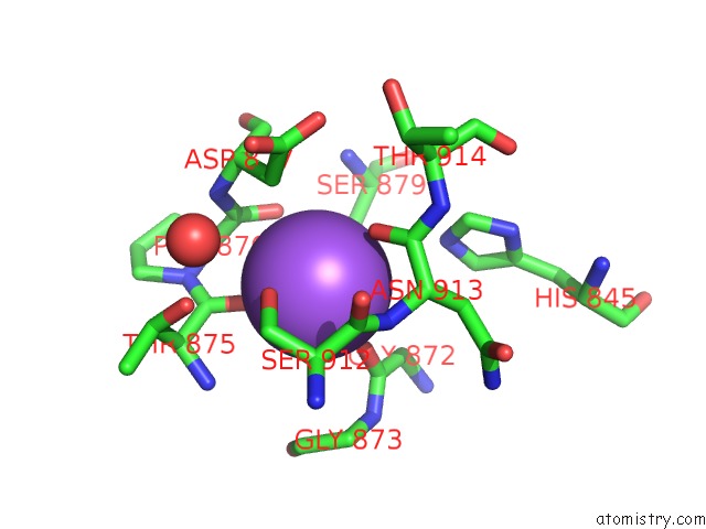
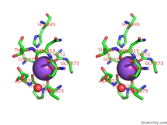
Sodium binding site 1 out of 4 in 3zyv
Go back to
Sodium binding site 1 out
of 4 in the Crystal Structure of the Mouse Liver Aldehyde Oxidase 3 (MAOX3)
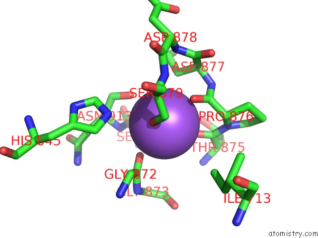
Mono view
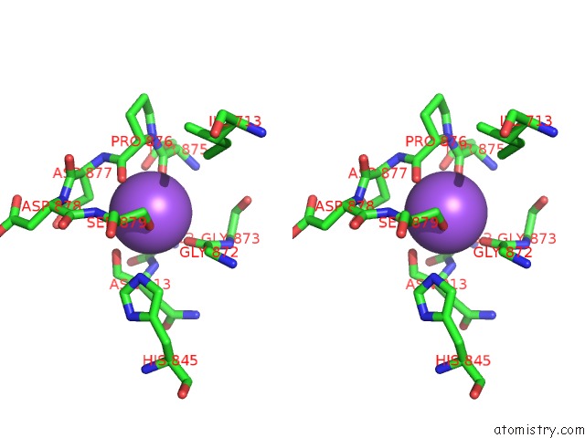
Stereo pair view
Mono view
Stereo pair view
A full contact list of Sodium with other atoms in the Na binding
site number 1 of Crystal Structure of the Mouse Liver Aldehyde Oxidase 3 (MAOX3) within 5.0Å range:
|
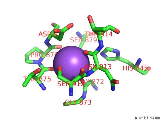
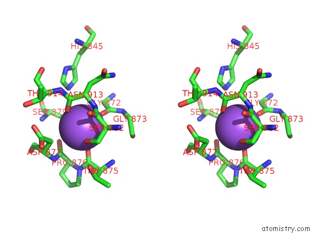
Sodium binding site 2 out of 4 in 3zyv
Go back to
Sodium binding site 2 out
of 4 in the Crystal Structure of the Mouse Liver Aldehyde Oxidase 3 (MAOX3)
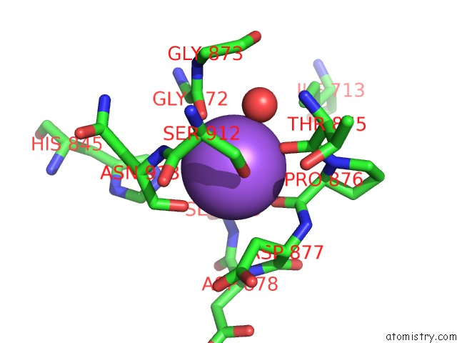
Mono view
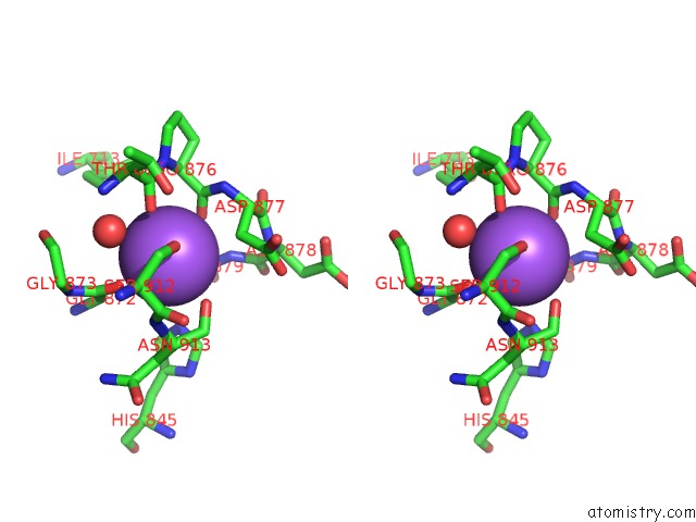
Stereo pair view
Mono view
Stereo pair view
A full contact list of Sodium with other atoms in the Na binding
site number 2 of Crystal Structure of the Mouse Liver Aldehyde Oxidase 3 (MAOX3) within 5.0Å range:
|
Sodium binding site 3 out of 4 in 3zyv
Go back to
Sodium binding site 3 out
of 4 in the Crystal Structure of the Mouse Liver Aldehyde Oxidase 3 (MAOX3)
Mono view
Stereo pair view
Mono view
Stereo pair view
A full contact list of Sodium with other atoms in the Na binding
site number 3 of Crystal Structure of the Mouse Liver Aldehyde Oxidase 3 (MAOX3) within 5.0Å range:
|
Sodium binding site 4 out of 4 in 3zyv
Go back to
Sodium binding site 4 out
of 4 in the Crystal Structure of the Mouse Liver Aldehyde Oxidase 3 (MAOX3)
Mono view
Stereo pair view
Mono view
Stereo pair view
A full contact list of Sodium with other atoms in the Na binding
site number 4 of Crystal Structure of the Mouse Liver Aldehyde Oxidase 3 (MAOX3) within 5.0Å range:
|
Reference:
C.Coelho,
M.Mahro,
J.Trincao,
A.T.P.Carvalho,
M.J.Ramos,
M.Terao,
E.Garattini,
S.Leimkuhler,
M.J.Romao.
The First Mammalian Aldehyde Oxidase Crystal Structure: Insights Into Substrate Specificity. J.Biol.Chem. V. 287 40690 2012.
ISSN: ISSN 0021-9258
PubMed: 23019336
DOI: 10.1074/JBC.M112.390419
Page generated: Mon Oct 7 14:15:00 2024
ISSN: ISSN 0021-9258
PubMed: 23019336
DOI: 10.1074/JBC.M112.390419
Last articles
Zn in 9MJ5Zn in 9HNW
Zn in 9G0L
Zn in 9FNE
Zn in 9DZN
Zn in 9E0I
Zn in 9D32
Zn in 9DAK
Zn in 8ZXC
Zn in 8ZUF