Sodium »
PDB 3vif-3way »
3wav »
Sodium in PDB 3wav: Crystal Structure of Autotaxin in Complex with Compound 10
Enzymatic activity of Crystal Structure of Autotaxin in Complex with Compound 10
All present enzymatic activity of Crystal Structure of Autotaxin in Complex with Compound 10:
3.1.4.39;
3.1.4.39;
Protein crystallography data
The structure of Crystal Structure of Autotaxin in Complex with Compound 10, PDB code: 3wav
was solved by
H.Nishimasu,
R.Ishitani,
O.Nureki,
with X-Ray Crystallography technique. A brief refinement statistics is given in the table below:
| Resolution Low / High (Å) | 33.24 / 1.80 |
| Space group | P 1 21 1 |
| Cell size a, b, c (Å), α, β, γ (°) | 61.505, 94.015, 75.446, 90.00, 94.52, 90.00 |
| R / Rfree (%) | 18.8 / 22.3 |
Other elements in 3wav:
The structure of Crystal Structure of Autotaxin in Complex with Compound 10 also contains other interesting chemical elements:
| Potassium | (K) | 1 atom |
| Zinc | (Zn) | 2 atoms |
| Calcium | (Ca) | 1 atom |
| Chlorine | (Cl) | 2 atoms |
Sodium Binding Sites:
The binding sites of Sodium atom in the Crystal Structure of Autotaxin in Complex with Compound 10
(pdb code 3wav). This binding sites where shown within
5.0 Angstroms radius around Sodium atom.
In total only one binding site of Sodium was determined in the Crystal Structure of Autotaxin in Complex with Compound 10, PDB code: 3wav:
In total only one binding site of Sodium was determined in the Crystal Structure of Autotaxin in Complex with Compound 10, PDB code: 3wav:
Sodium binding site 1 out of 1 in 3wav
Go back to
Sodium binding site 1 out
of 1 in the Crystal Structure of Autotaxin in Complex with Compound 10
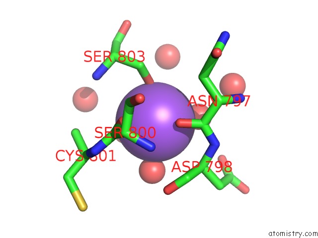
Mono view
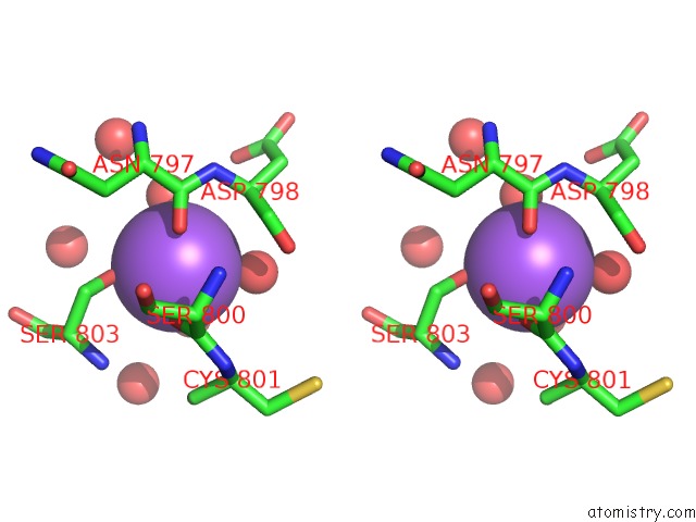
Stereo pair view
Mono view
Stereo pair view
A full contact list of Sodium with other atoms in the Na binding
site number 1 of Crystal Structure of Autotaxin in Complex with Compound 10 within 5.0Å range:
|
Reference:
M.Kawaguchi,
T.Okabe,
S.Okudaira,
H.Nishimasu,
R.Ishitani,
H.Kojima,
O.Nureki,
J.Aoki,
T.Nagano.
Screening and X-Ray Crystal Structure-Based Optimization of Autotaxin (ENPP2) Inhibitors, Using A Newly Developed Fluorescence Probe Acs Chem.Biol. V. 8 1713 2013.
ISSN: ISSN 1554-8929
PubMed: 23688339
DOI: 10.1021/CB400150C
Page generated: Mon Oct 7 13:55:25 2024
ISSN: ISSN 1554-8929
PubMed: 23688339
DOI: 10.1021/CB400150C
Last articles
Zn in 9MJ5Zn in 9HNW
Zn in 9G0L
Zn in 9FNE
Zn in 9DZN
Zn in 9E0I
Zn in 9D32
Zn in 9DAK
Zn in 8ZXC
Zn in 8ZUF