Sodium »
PDB 3vif-3way »
3w5c »
Sodium in PDB 3w5c: Crystal Structure of the Calcium Pump in the E2 State Free From Exogenous Inhibitors
Protein crystallography data
The structure of Crystal Structure of the Calcium Pump in the E2 State Free From Exogenous Inhibitors, PDB code: 3w5c
was solved by
C.Toyoshima,
S.Iwasawa,
H.Ogawa,
A.Hirata,
J.Tsueda,
G.Inesi,
with X-Ray Crystallography technique. A brief refinement statistics is given in the table below:
| Resolution Low / High (Å) | 15.00 / 2.50 |
| Space group | P 41 21 2 |
| Cell size a, b, c (Å), α, β, γ (°) | 71.226, 71.226, 586.236, 90.00, 90.00, 90.00 |
| R / Rfree (%) | 27 / 27.6 |
Sodium Binding Sites:
The binding sites of Sodium atom in the Crystal Structure of the Calcium Pump in the E2 State Free From Exogenous Inhibitors
(pdb code 3w5c). This binding sites where shown within
5.0 Angstroms radius around Sodium atom.
In total only one binding site of Sodium was determined in the Crystal Structure of the Calcium Pump in the E2 State Free From Exogenous Inhibitors, PDB code: 3w5c:
In total only one binding site of Sodium was determined in the Crystal Structure of the Calcium Pump in the E2 State Free From Exogenous Inhibitors, PDB code: 3w5c:
Sodium binding site 1 out of 1 in 3w5c
Go back to
Sodium binding site 1 out
of 1 in the Crystal Structure of the Calcium Pump in the E2 State Free From Exogenous Inhibitors
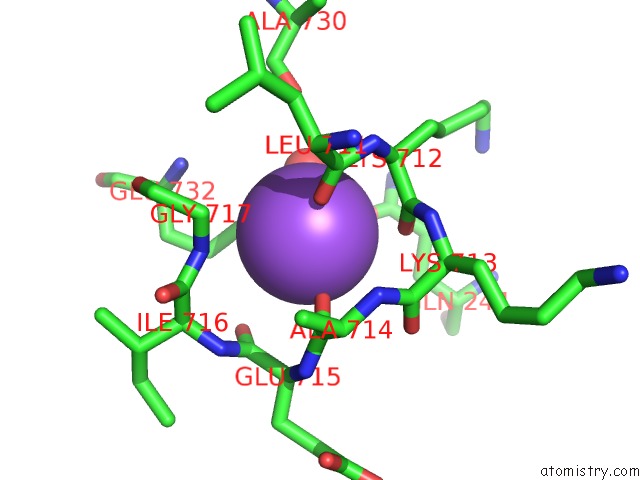
Mono view
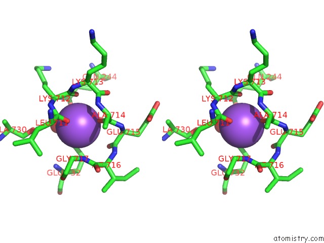
Stereo pair view
Mono view
Stereo pair view
A full contact list of Sodium with other atoms in the Na binding
site number 1 of Crystal Structure of the Calcium Pump in the E2 State Free From Exogenous Inhibitors within 5.0Å range:
|
Reference:
C.Toyoshima,
S.Iwasawa,
H.Ogawa,
A.Hirata,
J.Tsueda,
G.Inesi.
Crystal Structures of the Calcium Pump and Sarcolipin in the MG2+-Bound E1 State. Nature V. 495 260 2013.
ISSN: ISSN 0028-0836
PubMed: 23455422
DOI: 10.1038/NATURE11899
Page generated: Mon Oct 7 13:53:39 2024
ISSN: ISSN 0028-0836
PubMed: 23455422
DOI: 10.1038/NATURE11899
Last articles
Zn in 9J0NZn in 9J0O
Zn in 9J0P
Zn in 9FJX
Zn in 9EKB
Zn in 9C0F
Zn in 9CAH
Zn in 9CH0
Zn in 9CH3
Zn in 9CH1