Sodium »
PDB 3uq0-3v3r »
3usj »
Sodium in PDB 3usj: Crystal Structure of Leut Bound to L-Leucine in Space Group P21 From Lipid Bicelles
Protein crystallography data
The structure of Crystal Structure of Leut Bound to L-Leucine in Space Group P21 From Lipid Bicelles, PDB code: 3usj
was solved by
H.Wang,
J.Elferich,
E.Gouaux,
with X-Ray Crystallography technique. A brief refinement statistics is given in the table below:
| Resolution Low / High (Å) | 48.33 / 3.50 |
| Space group | P 1 21 1 |
| Cell size a, b, c (Å), α, β, γ (°) | 57.307, 179.880, 57.207, 90.00, 89.96, 90.00 |
| R / Rfree (%) | 26.4 / 30.9 |
Sodium Binding Sites:
The binding sites of Sodium atom in the Crystal Structure of Leut Bound to L-Leucine in Space Group P21 From Lipid Bicelles
(pdb code 3usj). This binding sites where shown within
5.0 Angstroms radius around Sodium atom.
In total 4 binding sites of Sodium where determined in the Crystal Structure of Leut Bound to L-Leucine in Space Group P21 From Lipid Bicelles, PDB code: 3usj:
Jump to Sodium binding site number: 1; 2; 3; 4;
In total 4 binding sites of Sodium where determined in the Crystal Structure of Leut Bound to L-Leucine in Space Group P21 From Lipid Bicelles, PDB code: 3usj:
Jump to Sodium binding site number: 1; 2; 3; 4;
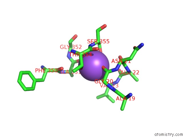
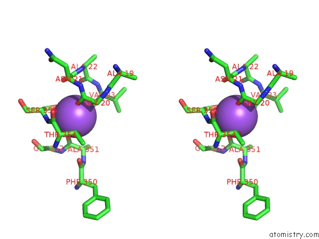
Sodium binding site 1 out of 4 in 3usj
Go back to
Sodium binding site 1 out
of 4 in the Crystal Structure of Leut Bound to L-Leucine in Space Group P21 From Lipid Bicelles
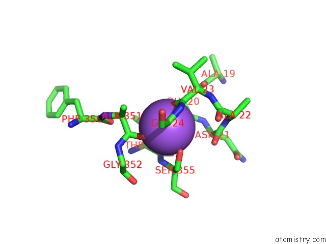
Mono view
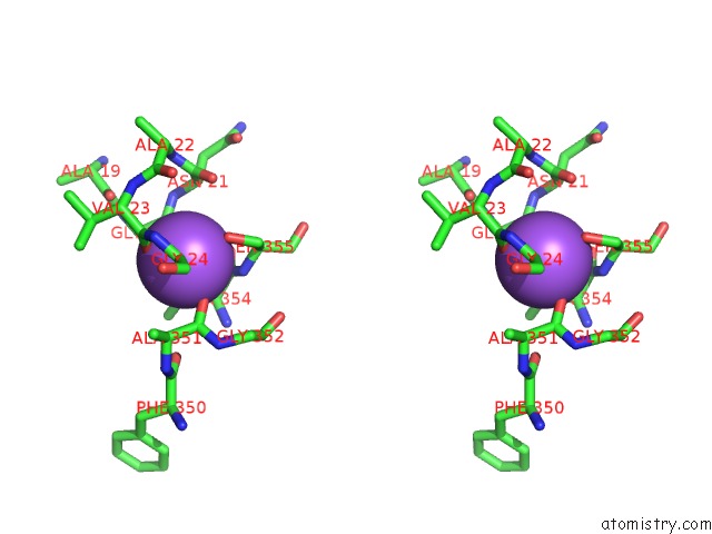
Stereo pair view
Mono view
Stereo pair view
A full contact list of Sodium with other atoms in the Na binding
site number 1 of Crystal Structure of Leut Bound to L-Leucine in Space Group P21 From Lipid Bicelles within 5.0Å range:
|
Sodium binding site 2 out of 4 in 3usj
Go back to
Sodium binding site 2 out
of 4 in the Crystal Structure of Leut Bound to L-Leucine in Space Group P21 From Lipid Bicelles
Mono view
Stereo pair view
Mono view
Stereo pair view
A full contact list of Sodium with other atoms in the Na binding
site number 2 of Crystal Structure of Leut Bound to L-Leucine in Space Group P21 From Lipid Bicelles within 5.0Å range:
|
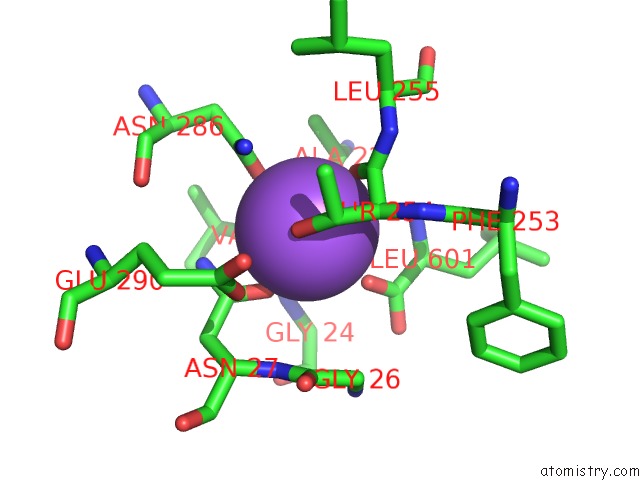
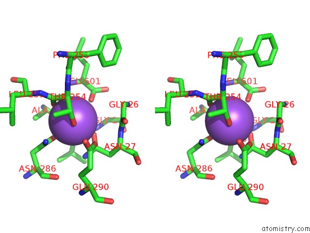
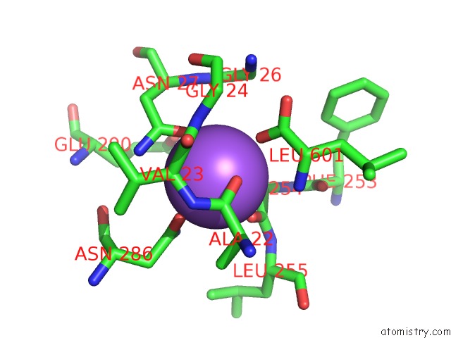
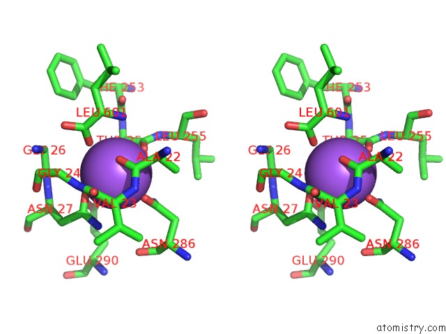
Sodium binding site 3 out of 4 in 3usj
Go back to
Sodium binding site 3 out
of 4 in the Crystal Structure of Leut Bound to L-Leucine in Space Group P21 From Lipid Bicelles
Mono view
Stereo pair view
Mono view
Stereo pair view
A full contact list of Sodium with other atoms in the Na binding
site number 3 of Crystal Structure of Leut Bound to L-Leucine in Space Group P21 From Lipid Bicelles within 5.0Å range:
|
Sodium binding site 4 out of 4 in 3usj
Go back to
Sodium binding site 4 out
of 4 in the Crystal Structure of Leut Bound to L-Leucine in Space Group P21 From Lipid Bicelles
Mono view
Stereo pair view
Mono view
Stereo pair view
A full contact list of Sodium with other atoms in the Na binding
site number 4 of Crystal Structure of Leut Bound to L-Leucine in Space Group P21 From Lipid Bicelles within 5.0Å range:
|
Reference:
H.Wang,
J.Elferich,
E.Gouaux.
Structures of Leut in Bicelles Define Conformation and Substrate Binding in A Membrane-Like Context. Nat.Struct.Mol.Biol. V. 19 212 2012.
ISSN: ISSN 1545-9993
PubMed: 22245965
DOI: 10.1038/NSMB.2215
Page generated: Mon Oct 7 13:32:02 2024
ISSN: ISSN 1545-9993
PubMed: 22245965
DOI: 10.1038/NSMB.2215
Last articles
Zn in 9MJ5Zn in 9HNW
Zn in 9G0L
Zn in 9FNE
Zn in 9DZN
Zn in 9E0I
Zn in 9D32
Zn in 9DAK
Zn in 8ZXC
Zn in 8ZUF