Sodium »
PDB 3tkj-3tzl »
3tkw »
Sodium in PDB 3tkw: Crystal Structure of Hiv Protease Model Precursor/Darunavir Complex
Enzymatic activity of Crystal Structure of Hiv Protease Model Precursor/Darunavir Complex
All present enzymatic activity of Crystal Structure of Hiv Protease Model Precursor/Darunavir Complex:
3.4.23.16;
3.4.23.16;
Protein crystallography data
The structure of Crystal Structure of Hiv Protease Model Precursor/Darunavir Complex, PDB code: 3tkw
was solved by
J.Agniswamy,
J.Sayer,
I.Weber,
J.Louis,
with X-Ray Crystallography technique. A brief refinement statistics is given in the table below:
| Resolution Low / High (Å) | 10.00 / 1.55 |
| Space group | P 21 21 2 |
| Cell size a, b, c (Å), α, β, γ (°) | 59.317, 86.223, 45.951, 90.00, 90.00, 90.00 |
| R / Rfree (%) | 16.7 / 23.1 |
Other elements in 3tkw:
The structure of Crystal Structure of Hiv Protease Model Precursor/Darunavir Complex also contains other interesting chemical elements:
| Chlorine | (Cl) | 3 atoms |
Sodium Binding Sites:
The binding sites of Sodium atom in the Crystal Structure of Hiv Protease Model Precursor/Darunavir Complex
(pdb code 3tkw). This binding sites where shown within
5.0 Angstroms radius around Sodium atom.
In total only one binding site of Sodium was determined in the Crystal Structure of Hiv Protease Model Precursor/Darunavir Complex, PDB code: 3tkw:
In total only one binding site of Sodium was determined in the Crystal Structure of Hiv Protease Model Precursor/Darunavir Complex, PDB code: 3tkw:
Sodium binding site 1 out of 1 in 3tkw
Go back to
Sodium binding site 1 out
of 1 in the Crystal Structure of Hiv Protease Model Precursor/Darunavir Complex
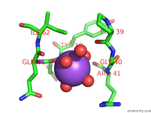
Mono view
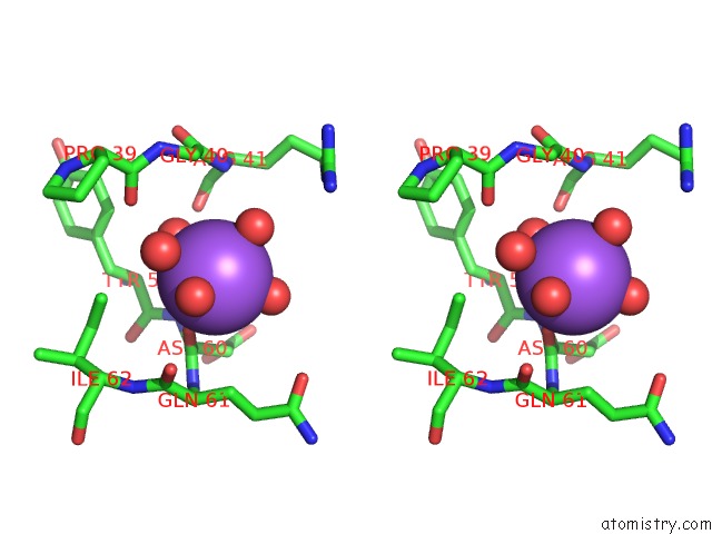
Stereo pair view
Mono view
Stereo pair view
A full contact list of Sodium with other atoms in the Na binding
site number 1 of Crystal Structure of Hiv Protease Model Precursor/Darunavir Complex within 5.0Å range:
|
Reference:
J.Agniswamy,
J.M.Sayer,
I.T.Weber,
J.M.Louis.
Terminal Interface Conformations Modulate Dimer Stability Prior to Amino Terminal Autoprocessing of Hiv-1 Protease. Biochemistry V. 51 1041 2012.
ISSN: ISSN 0006-2960
PubMed: 22242794
DOI: 10.1021/BI201809S
Page generated: Mon Oct 7 13:18:41 2024
ISSN: ISSN 0006-2960
PubMed: 22242794
DOI: 10.1021/BI201809S
Last articles
Ca in 5MLHCa in 5ML7
Ca in 5MKF
Ca in 5MKC
Ca in 5MIN
Ca in 5MKE
Ca in 5MJL
Ca in 5MIM
Ca in 5MIH
Ca in 5MJ7