Sodium »
PDB 3q11-3qpz »
3qjx »
Sodium in PDB 3qjx: Crystal Structure of E. Coli Aminopeptidase N in Complex with L-Serine
Enzymatic activity of Crystal Structure of E. Coli Aminopeptidase N in Complex with L-Serine
All present enzymatic activity of Crystal Structure of E. Coli Aminopeptidase N in Complex with L-Serine:
3.4.11.2;
3.4.11.2;
Protein crystallography data
The structure of Crystal Structure of E. Coli Aminopeptidase N in Complex with L-Serine, PDB code: 3qjx
was solved by
A.Addlagatta,
R.Gumpena,
C.Kishor,
R.J.Ganji,
with X-Ray Crystallography technique. A brief refinement statistics is given in the table below:
| Resolution Low / High (Å) | 35.74 / 1.45 |
| Space group | P 31 2 1 |
| Cell size a, b, c (Å), α, β, γ (°) | 120.230, 120.230, 170.801, 90.00, 90.00, 120.00 |
| R / Rfree (%) | 12 / 14.7 |
Other elements in 3qjx:
The structure of Crystal Structure of E. Coli Aminopeptidase N in Complex with L-Serine also contains other interesting chemical elements:
| Zinc | (Zn) | 1 atom |
Sodium Binding Sites:
The binding sites of Sodium atom in the Crystal Structure of E. Coli Aminopeptidase N in Complex with L-Serine
(pdb code 3qjx). This binding sites where shown within
5.0 Angstroms radius around Sodium atom.
In total 3 binding sites of Sodium where determined in the Crystal Structure of E. Coli Aminopeptidase N in Complex with L-Serine, PDB code: 3qjx:
Jump to Sodium binding site number: 1; 2; 3;
In total 3 binding sites of Sodium where determined in the Crystal Structure of E. Coli Aminopeptidase N in Complex with L-Serine, PDB code: 3qjx:
Jump to Sodium binding site number: 1; 2; 3;
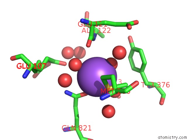
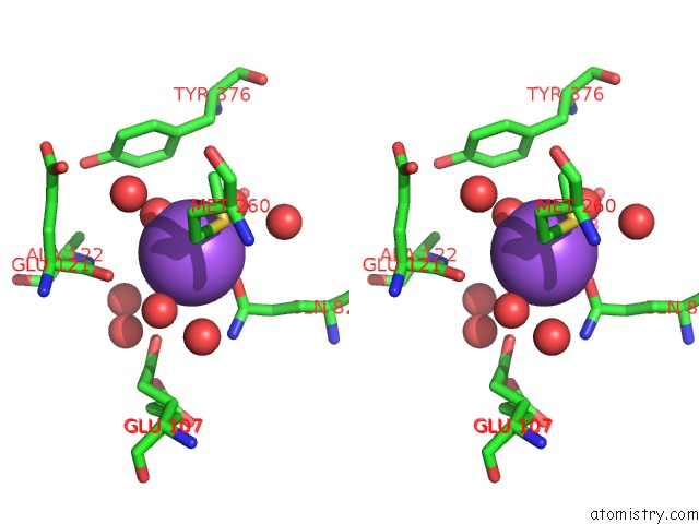
Sodium binding site 1 out of 3 in 3qjx
Go back to
Sodium binding site 1 out
of 3 in the Crystal Structure of E. Coli Aminopeptidase N in Complex with L-Serine
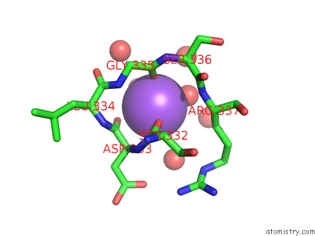
Mono view
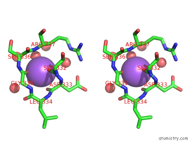
Stereo pair view
Mono view
Stereo pair view
A full contact list of Sodium with other atoms in the Na binding
site number 1 of Crystal Structure of E. Coli Aminopeptidase N in Complex with L-Serine within 5.0Å range:
|
Sodium binding site 2 out of 3 in 3qjx
Go back to
Sodium binding site 2 out
of 3 in the Crystal Structure of E. Coli Aminopeptidase N in Complex with L-Serine
Mono view
Stereo pair view
Mono view
Stereo pair view
A full contact list of Sodium with other atoms in the Na binding
site number 2 of Crystal Structure of E. Coli Aminopeptidase N in Complex with L-Serine within 5.0Å range:
|
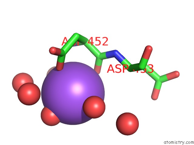
Sodium binding site 3 out of 3 in 3qjx
Go back to
Sodium binding site 3 out
of 3 in the Crystal Structure of E. Coli Aminopeptidase N in Complex with L-Serine
Mono view
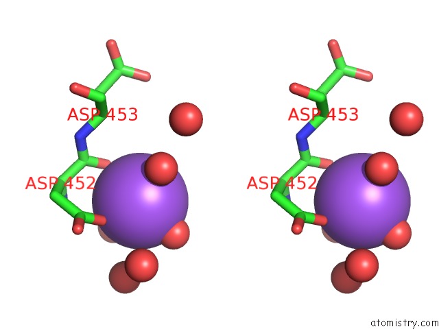
Stereo pair view
Mono view
Stereo pair view
A full contact list of Sodium with other atoms in the Na binding
site number 3 of Crystal Structure of E. Coli Aminopeptidase N in Complex with L-Serine within 5.0Å range:
|
Reference:
R.Gumpena,
C.Kishor,
R.J.Ganji,
A.Addlagatta.
Discovery of Alpha, Beta- and Alpha, Gamma-Diamino Acid Scaffolds For the Inhibition of M1 Family Aminopeptidases Chemmedchem V. 6 1971 2011.
ISSN: ISSN 1860-7179
PubMed: 22025387
DOI: 10.1002/CMDC.201100298
Page generated: Mon Oct 7 12:33:45 2024
ISSN: ISSN 1860-7179
PubMed: 22025387
DOI: 10.1002/CMDC.201100298
Last articles
Zn in 9J0NZn in 9J0O
Zn in 9J0P
Zn in 9FJX
Zn in 9EKB
Zn in 9C0F
Zn in 9CAH
Zn in 9CH0
Zn in 9CH3
Zn in 9CH1