Sodium »
PDB 3p2y-3pkh »
3pc4 »
Sodium in PDB 3pc4: Full Length Structure of Cystathionine Beta-Synthase From Drosophila in Complex with Serine
Enzymatic activity of Full Length Structure of Cystathionine Beta-Synthase From Drosophila in Complex with Serine
All present enzymatic activity of Full Length Structure of Cystathionine Beta-Synthase From Drosophila in Complex with Serine:
4.2.1.22;
4.2.1.22;
Protein crystallography data
The structure of Full Length Structure of Cystathionine Beta-Synthase From Drosophila in Complex with Serine, PDB code: 3pc4
was solved by
M.Koutmos,
J.L.Smith,
with X-Ray Crystallography technique. A brief refinement statistics is given in the table below:
| Resolution Low / High (Å) | 41.21 / 1.70 |
| Space group | C 2 2 21 |
| Cell size a, b, c (Å), α, β, γ (°) | 92.947, 137.935, 75.129, 90.00, 90.00, 90.00 |
| R / Rfree (%) | 17.9 / 20.3 |
Other elements in 3pc4:
The structure of Full Length Structure of Cystathionine Beta-Synthase From Drosophila in Complex with Serine also contains other interesting chemical elements:
| Iron | (Fe) | 1 atom |
Sodium Binding Sites:
The binding sites of Sodium atom in the Full Length Structure of Cystathionine Beta-Synthase From Drosophila in Complex with Serine
(pdb code 3pc4). This binding sites where shown within
5.0 Angstroms radius around Sodium atom.
In total only one binding site of Sodium was determined in the Full Length Structure of Cystathionine Beta-Synthase From Drosophila in Complex with Serine, PDB code: 3pc4:
In total only one binding site of Sodium was determined in the Full Length Structure of Cystathionine Beta-Synthase From Drosophila in Complex with Serine, PDB code: 3pc4:
Sodium binding site 1 out of 1 in 3pc4
Go back to
Sodium binding site 1 out
of 1 in the Full Length Structure of Cystathionine Beta-Synthase From Drosophila in Complex with Serine
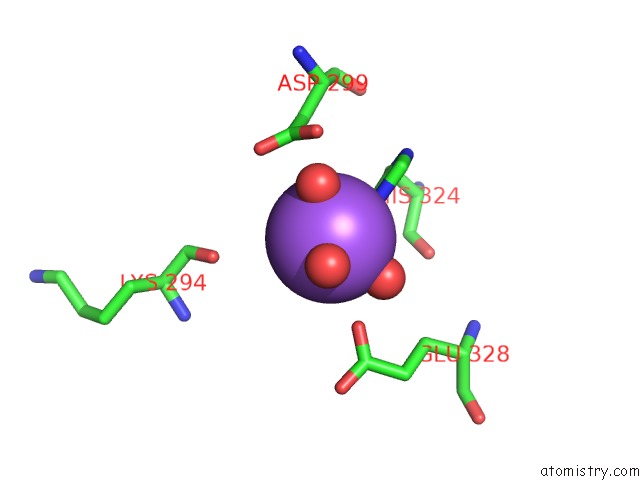
Mono view
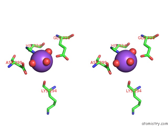
Stereo pair view
Mono view
Stereo pair view
A full contact list of Sodium with other atoms in the Na binding
site number 1 of Full Length Structure of Cystathionine Beta-Synthase From Drosophila in Complex with Serine within 5.0Å range:
|
Reference:
M.Koutmos,
O.Kabil,
J.L.Smith,
R.Banerjee.
Structural Basis For Substrate Activation and Regulation By Cystathionine Beta-Synthase (Cbs) Domains in Cystathionine {Beta}-Synthase. Proc.Natl.Acad.Sci.Usa V. 107 20958 2010.
ISSN: ISSN 0027-8424
PubMed: 21081698
DOI: 10.1073/PNAS.1011448107
Page generated: Mon Oct 7 12:12:32 2024
ISSN: ISSN 0027-8424
PubMed: 21081698
DOI: 10.1073/PNAS.1011448107
Last articles
F in 7N2AF in 7MXY
F in 7MYY
F in 7N13
F in 7MYU
F in 7MYR
F in 7MYO
F in 7MXN
F in 7MXG
F in 7MXH