Sodium »
PDB 3ogc-3p1l »
3oyo »
Sodium in PDB 3oyo: Crystal Structure of Hemopexin Fold Protein CP4 From Cow Pea
Protein crystallography data
The structure of Crystal Structure of Hemopexin Fold Protein CP4 From Cow Pea, PDB code: 3oyo
was solved by
V.Gaur,
V.Chanana,
D.M.Salunke,
with X-Ray Crystallography technique. A brief refinement statistics is given in the table below:
| Resolution Low / High (Å) | 26.22 / 2.10 |
| Space group | C 1 2 1 |
| Cell size a, b, c (Å), α, β, γ (°) | 125.087, 60.138, 67.574, 90.00, 111.18, 90.00 |
| R / Rfree (%) | 20.5 / 24.6 |
Other elements in 3oyo:
The structure of Crystal Structure of Hemopexin Fold Protein CP4 From Cow Pea also contains other interesting chemical elements:
| Chlorine | (Cl) | 2 atoms |
| Calcium | (Ca) | 2 atoms |
Sodium Binding Sites:
The binding sites of Sodium atom in the Crystal Structure of Hemopexin Fold Protein CP4 From Cow Pea
(pdb code 3oyo). This binding sites where shown within
5.0 Angstroms radius around Sodium atom.
In total 2 binding sites of Sodium where determined in the Crystal Structure of Hemopexin Fold Protein CP4 From Cow Pea, PDB code: 3oyo:
Jump to Sodium binding site number: 1; 2;
In total 2 binding sites of Sodium where determined in the Crystal Structure of Hemopexin Fold Protein CP4 From Cow Pea, PDB code: 3oyo:
Jump to Sodium binding site number: 1; 2;
Sodium binding site 1 out of 2 in 3oyo
Go back to
Sodium binding site 1 out
of 2 in the Crystal Structure of Hemopexin Fold Protein CP4 From Cow Pea
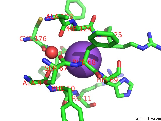
Mono view
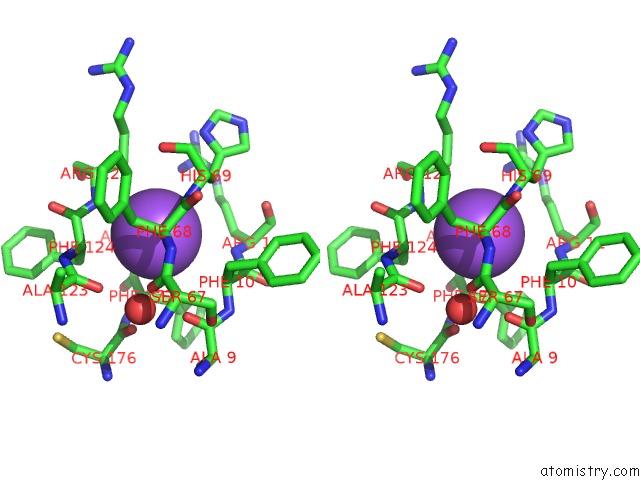
Stereo pair view
Mono view
Stereo pair view
A full contact list of Sodium with other atoms in the Na binding
site number 1 of Crystal Structure of Hemopexin Fold Protein CP4 From Cow Pea within 5.0Å range:
|
Sodium binding site 2 out of 2 in 3oyo
Go back to
Sodium binding site 2 out
of 2 in the Crystal Structure of Hemopexin Fold Protein CP4 From Cow Pea
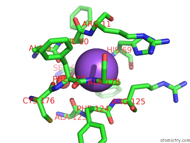
Mono view
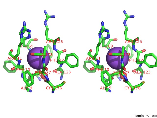
Stereo pair view
Mono view
Stereo pair view
A full contact list of Sodium with other atoms in the Na binding
site number 2 of Crystal Structure of Hemopexin Fold Protein CP4 From Cow Pea within 5.0Å range:
|
Reference:
V.Gaur,
V.Chanana,
A.Jain,
D.M.Salunke.
The Structure of A Haemopexin-Fold Protein From Cow Pea (Vigna Unguiculata) Suggests Functional Diversity of Haemopexins in Plants Acta Crystallogr.,Sect.F V. 67 193 2011.
ISSN: ESSN 1744-3091
PubMed: 21301085
DOI: 10.1107/S1744309110051250
Page generated: Mon Oct 7 12:07:42 2024
ISSN: ESSN 1744-3091
PubMed: 21301085
DOI: 10.1107/S1744309110051250
Last articles
Ca in 5NEMCa in 5NE5
Ca in 5NBP
Ca in 5NBN
Ca in 5NBM
Ca in 5NBL
Ca in 5N7G
Ca in 5N7F
Ca in 5N7D
Ca in 5N5P