Sodium »
PDB 3ogc-3p1l »
3okt »
Sodium in PDB 3okt: Mouse Plexin A2, Extracellular Domains 1-4
Protein crystallography data
The structure of Mouse Plexin A2, Extracellular Domains 1-4, PDB code: 3okt
was solved by
B.J.C.Janssen,
R.A.Robinson,
C.H.Bell,
C.Siebold,
E.Y.Jones,
with X-Ray Crystallography technique. A brief refinement statistics is given in the table below:
| Resolution Low / High (Å) | 44.91 / 2.30 |
| Space group | C 1 2 1 |
| Cell size a, b, c (Å), α, β, γ (°) | 157.860, 92.903, 62.904, 90.00, 102.87, 90.00 |
| R / Rfree (%) | 19.6 / 25.1 |
Other elements in 3okt:
The structure of Mouse Plexin A2, Extracellular Domains 1-4 also contains other interesting chemical elements:
| Chlorine | (Cl) | 2 atoms |
Sodium Binding Sites:
The binding sites of Sodium atom in the Mouse Plexin A2, Extracellular Domains 1-4
(pdb code 3okt). This binding sites where shown within
5.0 Angstroms radius around Sodium atom.
In total only one binding site of Sodium was determined in the Mouse Plexin A2, Extracellular Domains 1-4, PDB code: 3okt:
In total only one binding site of Sodium was determined in the Mouse Plexin A2, Extracellular Domains 1-4, PDB code: 3okt:
Sodium binding site 1 out of 1 in 3okt
Go back to
Sodium binding site 1 out
of 1 in the Mouse Plexin A2, Extracellular Domains 1-4
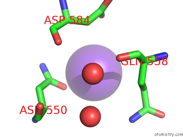
Mono view
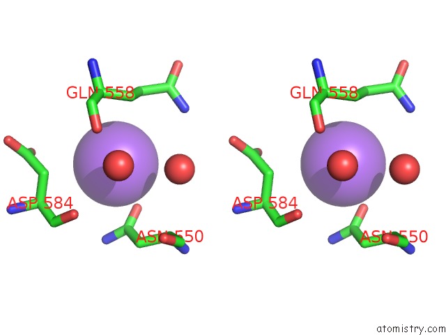
Stereo pair view
Mono view
Stereo pair view
A full contact list of Sodium with other atoms in the Na binding
site number 1 of Mouse Plexin A2, Extracellular Domains 1-4 within 5.0Å range:
|
Reference:
B.J.Janssen,
R.A.Robinson,
F.Perez-Branguli,
C.H.Bell,
K.J.Mitchell,
C.Siebold,
E.Y.Jones.
Structural Basis of Semaphorin-Plexin Signalling. Nature V. 467 1118 2010.
ISSN: ISSN 0028-0836
PubMed: 20877282
DOI: 10.1038/NATURE09468
Page generated: Mon Oct 7 12:03:14 2024
ISSN: ISSN 0028-0836
PubMed: 20877282
DOI: 10.1038/NATURE09468
Last articles
Ca in 5O25Ca in 5O1U
Ca in 5O0S
Ca in 5NZE
Ca in 5NZ4
Ca in 5NWE
Ca in 5NYY
Ca in 5NXL
Ca in 5NXU
Ca in 5NXR