Sodium »
PDB 3nte-3off »
3nyt »
Sodium in PDB 3nyt: X-Ray Crystal Structure of the Wlbe (Wpbe) Aminotransferase From Pseudomonas Aeruginosa, Mutation K185A, in Complex with the Plp External Aldimine Adduct with Udp-3-Amino-2-N-Acetyl-Glucuronic Acid, at 1.3 Angstrom Resolution
Protein crystallography data
The structure of X-Ray Crystal Structure of the Wlbe (Wpbe) Aminotransferase From Pseudomonas Aeruginosa, Mutation K185A, in Complex with the Plp External Aldimine Adduct with Udp-3-Amino-2-N-Acetyl-Glucuronic Acid, at 1.3 Angstrom Resolution, PDB code: 3nyt
was solved by
H.M.Holden,
J.B.Thoden,
with X-Ray Crystallography technique. A brief refinement statistics is given in the table below:
| Resolution Low / High (Å) | 30.00 / 1.30 |
| Space group | C 1 2 1 |
| Cell size a, b, c (Å), α, β, γ (°) | 59.684, 92.170, 75.652, 90.00, 111.84, 90.00 |
| R / Rfree (%) | 19 / 21.6 |
Sodium Binding Sites:
The binding sites of Sodium atom in the X-Ray Crystal Structure of the Wlbe (Wpbe) Aminotransferase From Pseudomonas Aeruginosa, Mutation K185A, in Complex with the Plp External Aldimine Adduct with Udp-3-Amino-2-N-Acetyl-Glucuronic Acid, at 1.3 Angstrom Resolution
(pdb code 3nyt). This binding sites where shown within
5.0 Angstroms radius around Sodium atom.
In total only one binding site of Sodium was determined in the X-Ray Crystal Structure of the Wlbe (Wpbe) Aminotransferase From Pseudomonas Aeruginosa, Mutation K185A, in Complex with the Plp External Aldimine Adduct with Udp-3-Amino-2-N-Acetyl-Glucuronic Acid, at 1.3 Angstrom Resolution, PDB code: 3nyt:
In total only one binding site of Sodium was determined in the X-Ray Crystal Structure of the Wlbe (Wpbe) Aminotransferase From Pseudomonas Aeruginosa, Mutation K185A, in Complex with the Plp External Aldimine Adduct with Udp-3-Amino-2-N-Acetyl-Glucuronic Acid, at 1.3 Angstrom Resolution, PDB code: 3nyt:
Sodium binding site 1 out of 1 in 3nyt
Go back to
Sodium binding site 1 out
of 1 in the X-Ray Crystal Structure of the Wlbe (Wpbe) Aminotransferase From Pseudomonas Aeruginosa, Mutation K185A, in Complex with the Plp External Aldimine Adduct with Udp-3-Amino-2-N-Acetyl-Glucuronic Acid, at 1.3 Angstrom Resolution
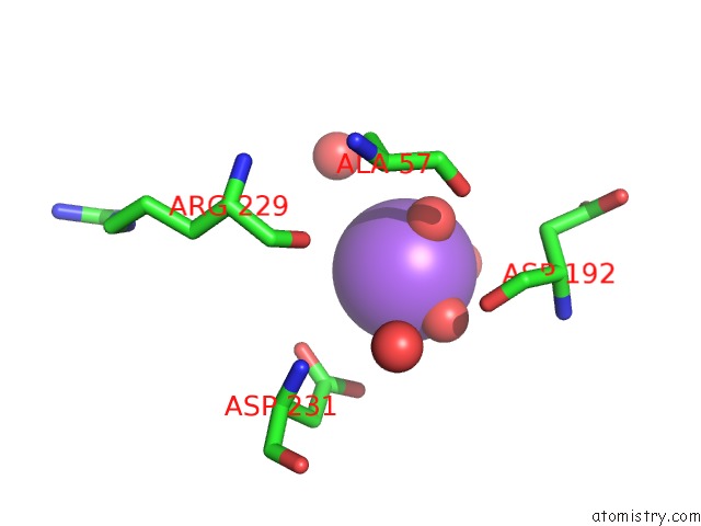
Mono view
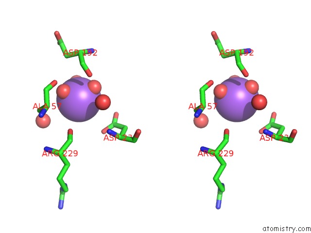
Stereo pair view
Mono view
Stereo pair view
A full contact list of Sodium with other atoms in the Na binding
site number 1 of X-Ray Crystal Structure of the Wlbe (Wpbe) Aminotransferase From Pseudomonas Aeruginosa, Mutation K185A, in Complex with the Plp External Aldimine Adduct with Udp-3-Amino-2-N-Acetyl-Glucuronic Acid, at 1.3 Angstrom Resolution within 5.0Å range:
|
Reference:
G.T.Dow,
M.Gilbert,
J.B.Thoden,
H.M.Holden.
Structural Investigation on Wlarg From Campylobacter Jejuni: A Sugar Aminotransferase. Protein Sci. V. 26 586 2017.
ISSN: ISSN 0961-8368
PubMed: 28028852
DOI: 10.1002/PRO.3109
Page generated: Mon Oct 7 11:55:27 2024
ISSN: ISSN 0961-8368
PubMed: 28028852
DOI: 10.1002/PRO.3109
Last articles
Cl in 5SYWCl in 5SYX
Cl in 5SYY
Cl in 5SYM
Cl in 5SYV
Cl in 5SYU
Cl in 5SYL
Cl in 5SYK
Cl in 5SYJ
Cl in 5SYI