Sodium in PDB 3ngj: Crystal Structure of A Putative Deoxyribose-Phosphate Aldolase From Entamoeba Histolytica
Protein crystallography data
The structure of Crystal Structure of A Putative Deoxyribose-Phosphate Aldolase From Entamoeba Histolytica, PDB code: 3ngj
was solved by
Seattle Structural Genomics Center For Infectious Disease (Ssgcid),
with X-Ray Crystallography technique. A brief refinement statistics is given in the table below:
Resolution Low / High (Å)
|
47.38 /
1.70
|
Space group
|
P 1 21 1
|
Cell size a, b, c (Å), α, β, γ (°)
|
50.260,
70.780,
126.750,
90.00,
92.46,
90.00
|
R / Rfree (%)
|
14.6 /
17.5
|
Other elements in 3ngj:
The structure of Crystal Structure of A Putative Deoxyribose-Phosphate Aldolase From Entamoeba Histolytica also contains other interesting chemical elements:
Sodium Binding Sites:
The binding sites of Sodium atom in the Crystal Structure of A Putative Deoxyribose-Phosphate Aldolase From Entamoeba Histolytica
(pdb code 3ngj). This binding sites where shown within
5.0 Angstroms radius around Sodium atom.
In total 4 binding sites of Sodium where determined in the
Crystal Structure of A Putative Deoxyribose-Phosphate Aldolase From Entamoeba Histolytica, PDB code: 3ngj:
Jump to Sodium binding site number:
1;
2;
3;
4;
Sodium binding site 1 out
of 4 in 3ngj
Go back to
Sodium Binding Sites List in 3ngj
Sodium binding site 1 out
of 4 in the Crystal Structure of A Putative Deoxyribose-Phosphate Aldolase From Entamoeba Histolytica
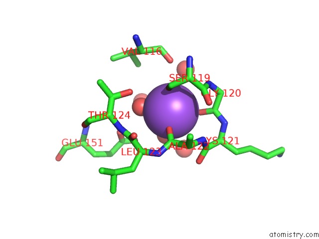
Mono view
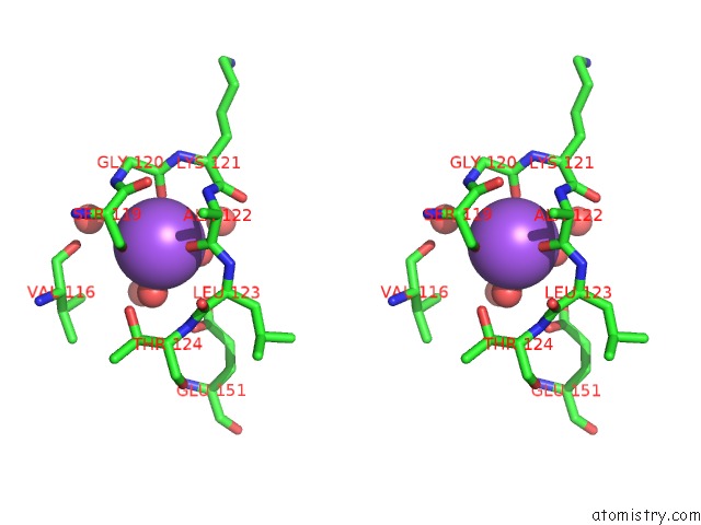
Stereo pair view
|
A full contact list of Sodium with other atoms in the Na binding
site number 1 of Crystal Structure of A Putative Deoxyribose-Phosphate Aldolase From Entamoeba Histolytica within 5.0Å range:
probe
|
atom
|
residue
|
distance (Å)
|
B
|
Occ
|
A:Na249
b:12.2
occ:1.00
|
O
|
A:ALA122
|
2.3
|
9.7
|
1.0
|
OG
|
A:SER119
|
2.4
|
9.1
|
1.0
|
O
|
A:GLY120
|
2.4
|
12.0
|
1.0
|
O
|
A:HOH276
|
2.4
|
22.8
|
1.0
|
O
|
A:HOH263
|
2.5
|
12.4
|
1.0
|
O
|
A:HOH306
|
2.6
|
14.8
|
1.0
|
C
|
A:ALA122
|
3.5
|
10.8
|
1.0
|
CB
|
A:SER119
|
3.5
|
9.1
|
1.0
|
C
|
A:GLY120
|
3.5
|
11.9
|
1.0
|
N
|
A:GLY120
|
3.9
|
10.2
|
1.0
|
C
|
A:SER119
|
3.9
|
9.2
|
1.0
|
O
|
A:HOH822
|
4.0
|
36.7
|
1.0
|
N
|
A:ALA122
|
4.1
|
11.1
|
1.0
|
O
|
A:VAL116
|
4.1
|
9.2
|
1.0
|
OG1
|
A:THR124
|
4.2
|
11.2
|
1.0
|
C
|
A:LYS121
|
4.2
|
12.9
|
1.0
|
O
|
A:SER119
|
4.2
|
9.8
|
1.0
|
CA
|
A:GLY120
|
4.3
|
11.3
|
1.0
|
CA
|
A:SER119
|
4.3
|
9.1
|
1.0
|
O
|
A:HOH818
|
4.3
|
32.1
|
1.0
|
CA
|
A:ALA122
|
4.4
|
10.3
|
1.0
|
O
|
A:HOH821
|
4.4
|
38.7
|
1.0
|
N
|
A:LEU123
|
4.4
|
10.5
|
1.0
|
N
|
A:LYS121
|
4.4
|
13.0
|
1.0
|
O
|
A:HOH593
|
4.5
|
26.9
|
1.0
|
OE2
|
A:GLU151
|
4.5
|
16.8
|
1.0
|
CA
|
A:LEU123
|
4.5
|
10.7
|
1.0
|
OE1
|
A:GLU151
|
4.5
|
9.9
|
1.0
|
CA
|
A:LYS121
|
4.6
|
13.8
|
1.0
|
O
|
A:LYS121
|
4.7
|
13.5
|
1.0
|
C
|
A:LEU123
|
4.8
|
10.2
|
1.0
|
N
|
A:THR124
|
4.8
|
9.6
|
1.0
|
CD
|
A:GLU151
|
5.0
|
13.1
|
1.0
|
|
Sodium binding site 2 out
of 4 in 3ngj
Go back to
Sodium Binding Sites List in 3ngj
Sodium binding site 2 out
of 4 in the Crystal Structure of A Putative Deoxyribose-Phosphate Aldolase From Entamoeba Histolytica
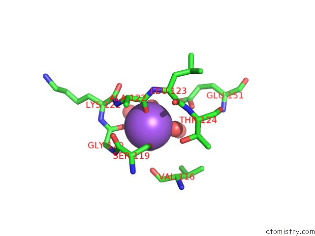
Mono view
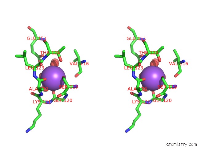
Stereo pair view
|
A full contact list of Sodium with other atoms in the Na binding
site number 2 of Crystal Structure of A Putative Deoxyribose-Phosphate Aldolase From Entamoeba Histolytica within 5.0Å range:
probe
|
atom
|
residue
|
distance (Å)
|
B
|
Occ
|
B:Na249
b:17.3
occ:1.00
|
O
|
B:ALA122
|
2.2
|
16.4
|
1.0
|
OG
|
B:SER119
|
2.3
|
14.6
|
1.0
|
O
|
B:HOH226
|
2.4
|
18.3
|
1.0
|
O
|
B:GLY120
|
2.5
|
18.6
|
1.0
|
O
|
B:HOH227
|
2.5
|
14.4
|
1.0
|
O
|
B:HOH355
|
2.5
|
23.9
|
1.0
|
CB
|
B:SER119
|
3.4
|
14.4
|
1.0
|
C
|
B:ALA122
|
3.4
|
16.5
|
1.0
|
C
|
B:GLY120
|
3.5
|
18.9
|
1.0
|
C
|
B:SER119
|
4.0
|
15.3
|
1.0
|
O
|
B:HOH780
|
4.0
|
41.9
|
1.0
|
N
|
B:GLY120
|
4.0
|
17.0
|
1.0
|
N
|
B:ALA122
|
4.0
|
18.0
|
1.0
|
O
|
B:VAL116
|
4.1
|
13.5
|
1.0
|
O
|
B:HOH847
|
4.2
|
25.5
|
1.0
|
OG1
|
B:THR124
|
4.2
|
14.2
|
1.0
|
C
|
B:LYS121
|
4.2
|
20.4
|
1.0
|
O
|
B:SER119
|
4.3
|
14.9
|
1.0
|
N
|
B:LEU123
|
4.3
|
15.6
|
1.0
|
CA
|
B:SER119
|
4.3
|
14.5
|
1.0
|
CA
|
B:LEU123
|
4.3
|
15.6
|
1.0
|
CA
|
B:GLY120
|
4.3
|
18.4
|
1.0
|
CA
|
B:ALA122
|
4.3
|
16.9
|
1.0
|
OE2
|
B:GLU151
|
4.4
|
17.7
|
1.0
|
OE1
|
B:GLU151
|
4.5
|
14.5
|
1.0
|
N
|
B:LYS121
|
4.5
|
20.5
|
1.0
|
N
|
B:THR124
|
4.6
|
13.5
|
1.0
|
C
|
B:LEU123
|
4.6
|
14.7
|
1.0
|
O
|
B:LYS121
|
4.6
|
20.4
|
1.0
|
CA
|
B:LYS121
|
4.7
|
21.3
|
1.0
|
CD
|
B:GLU151
|
4.8
|
16.6
|
1.0
|
|
Sodium binding site 3 out
of 4 in 3ngj
Go back to
Sodium Binding Sites List in 3ngj
Sodium binding site 3 out
of 4 in the Crystal Structure of A Putative Deoxyribose-Phosphate Aldolase From Entamoeba Histolytica
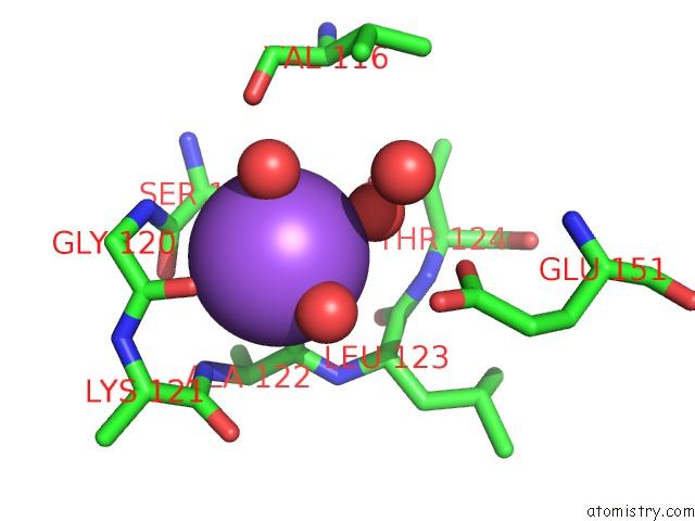
Mono view
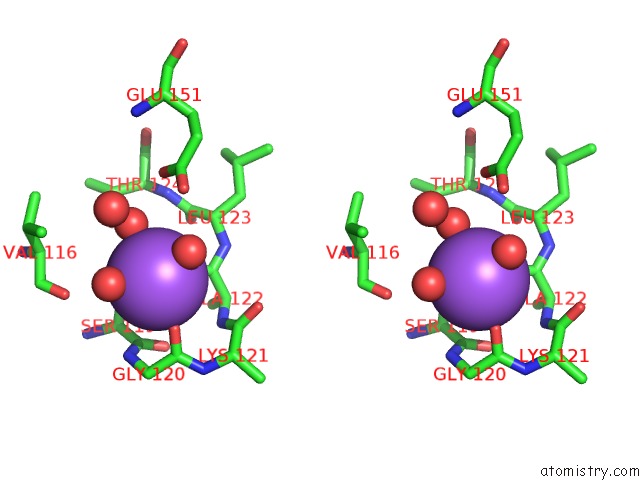
Stereo pair view
|
A full contact list of Sodium with other atoms in the Na binding
site number 3 of Crystal Structure of A Putative Deoxyribose-Phosphate Aldolase From Entamoeba Histolytica within 5.0Å range:
probe
|
atom
|
residue
|
distance (Å)
|
B
|
Occ
|
C:Na249
b:15.3
occ:1.00
|
O
|
C:ALA122
|
2.2
|
14.0
|
1.0
|
O
|
C:GLY120
|
2.3
|
16.4
|
1.0
|
OG
|
C:SER119
|
2.4
|
14.3
|
1.0
|
O
|
C:HOH318
|
2.4
|
18.4
|
1.0
|
O
|
C:HOH220
|
2.6
|
29.8
|
1.0
|
O
|
C:HOH240
|
2.7
|
14.4
|
1.0
|
C
|
C:ALA122
|
3.4
|
14.0
|
1.0
|
CB
|
C:SER119
|
3.4
|
14.1
|
1.0
|
C
|
C:GLY120
|
3.5
|
16.9
|
1.0
|
C
|
C:SER119
|
3.8
|
15.2
|
1.0
|
N
|
C:GLY120
|
3.8
|
16.2
|
1.0
|
N
|
C:ALA122
|
4.0
|
14.7
|
1.0
|
O
|
C:SER119
|
4.1
|
14.2
|
1.0
|
O
|
C:VAL116
|
4.2
|
14.9
|
1.0
|
CA
|
C:GLY120
|
4.2
|
17.3
|
1.0
|
CA
|
C:SER119
|
4.2
|
14.6
|
1.0
|
C
|
C:LYS121
|
4.2
|
16.1
|
1.0
|
N
|
C:LEU123
|
4.3
|
14.0
|
1.0
|
CA
|
C:ALA122
|
4.3
|
14.2
|
1.0
|
OG1
|
C:THR124
|
4.4
|
14.8
|
1.0
|
CA
|
C:LEU123
|
4.4
|
14.1
|
1.0
|
O
|
C:HOH436
|
4.4
|
26.2
|
1.0
|
N
|
C:LYS121
|
4.5
|
16.7
|
1.0
|
OE2
|
C:GLU151
|
4.5
|
18.5
|
1.0
|
O
|
C:LYS121
|
4.6
|
17.4
|
1.0
|
OE1
|
C:GLU151
|
4.7
|
13.6
|
1.0
|
CA
|
C:LYS121
|
4.7
|
17.4
|
1.0
|
C
|
C:LEU123
|
4.7
|
13.3
|
1.0
|
N
|
C:THR124
|
4.8
|
12.0
|
1.0
|
|
Sodium binding site 4 out
of 4 in 3ngj
Go back to
Sodium Binding Sites List in 3ngj
Sodium binding site 4 out
of 4 in the Crystal Structure of A Putative Deoxyribose-Phosphate Aldolase From Entamoeba Histolytica
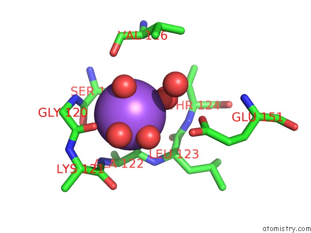
Mono view
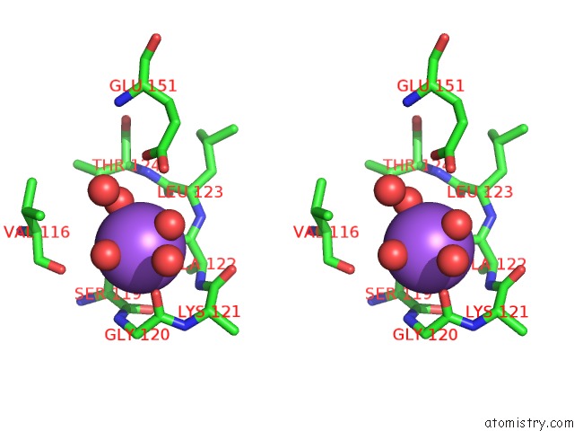
Stereo pair view
|
A full contact list of Sodium with other atoms in the Na binding
site number 4 of Crystal Structure of A Putative Deoxyribose-Phosphate Aldolase From Entamoeba Histolytica within 5.0Å range:
probe
|
atom
|
residue
|
distance (Å)
|
B
|
Occ
|
D:Na249
b:9.9
occ:1.00
|
O
|
D:ALA122
|
2.2
|
9.6
|
1.0
|
OG
|
D:SER119
|
2.3
|
6.6
|
1.0
|
O
|
D:HOH433
|
2.4
|
16.1
|
1.0
|
O
|
D:HOH462
|
2.4
|
17.2
|
1.0
|
O
|
D:GLY120
|
2.4
|
12.2
|
1.0
|
O
|
D:HOH220
|
2.5
|
10.3
|
1.0
|
C
|
D:ALA122
|
3.4
|
9.2
|
1.0
|
CB
|
D:SER119
|
3.5
|
8.6
|
1.0
|
C
|
D:GLY120
|
3.5
|
10.2
|
1.0
|
N
|
D:GLY120
|
3.9
|
7.7
|
1.0
|
C
|
D:SER119
|
3.9
|
7.7
|
1.0
|
O
|
D:HOH734
|
4.0
|
32.1
|
1.0
|
N
|
D:ALA122
|
4.0
|
9.6
|
1.0
|
O
|
D:VAL116
|
4.1
|
8.1
|
1.0
|
O
|
D:SER119
|
4.2
|
6.3
|
1.0
|
C
|
D:LYS121
|
4.2
|
11.9
|
1.0
|
OG1
|
D:THR124
|
4.3
|
9.2
|
1.0
|
CA
|
D:GLY120
|
4.3
|
10.2
|
1.0
|
N
|
D:LEU123
|
4.3
|
9.6
|
1.0
|
CA
|
D:SER119
|
4.3
|
6.4
|
1.0
|
CA
|
D:ALA122
|
4.3
|
9.7
|
1.0
|
O
|
D:HOH733
|
4.4
|
21.8
|
1.0
|
CA
|
D:LEU123
|
4.4
|
9.6
|
1.0
|
N
|
D:LYS121
|
4.4
|
11.7
|
1.0
|
OE2
|
D:GLU151
|
4.5
|
14.2
|
1.0
|
OE1
|
D:GLU151
|
4.6
|
8.0
|
1.0
|
CA
|
D:LYS121
|
4.6
|
12.4
|
1.0
|
O
|
D:LYS121
|
4.7
|
13.0
|
1.0
|
N
|
D:THR124
|
4.7
|
7.3
|
1.0
|
C
|
D:LEU123
|
4.7
|
8.4
|
1.0
|
CD
|
D:GLU151
|
5.0
|
8.2
|
1.0
|
|
Reference:
J.Abendroth,
A.Gardberg,
B.Staker.
Crystal Structure of A Putative Deoxyribose-Phosphate Aldolase From Entamoeba Histolytica To Be Published.
Page generated: Mon Oct 7 11:52:10 2024
|