Sodium »
PDB 3n0u-3nrv »
3n7z »
Sodium in PDB 3n7z: Crystal Structure of Acetyltransferase From Bacillus Anthracis
Protein crystallography data
The structure of Crystal Structure of Acetyltransferase From Bacillus Anthracis, PDB code: 3n7z
was solved by
C.Chang,
R.Wu,
P.Gornicki,
R.Zhang,
A.Joachimiak,
Midwest Center Forstructural Genomics (Mcsg),
with X-Ray Crystallography technique. A brief refinement statistics is given in the table below:
| Resolution Low / High (Å) | 50.00 / 2.75 |
| Space group | P 1 21 1 |
| Cell size a, b, c (Å), α, β, γ (°) | 79.429, 176.859, 109.973, 90.00, 105.73, 90.00 |
| R / Rfree (%) | 18.7 / 24.2 |
Sodium Binding Sites:
The binding sites of Sodium atom in the Crystal Structure of Acetyltransferase From Bacillus Anthracis
(pdb code 3n7z). This binding sites where shown within
5.0 Angstroms radius around Sodium atom.
In total 6 binding sites of Sodium where determined in the Crystal Structure of Acetyltransferase From Bacillus Anthracis, PDB code: 3n7z:
Jump to Sodium binding site number: 1; 2; 3; 4; 5; 6;
In total 6 binding sites of Sodium where determined in the Crystal Structure of Acetyltransferase From Bacillus Anthracis, PDB code: 3n7z:
Jump to Sodium binding site number: 1; 2; 3; 4; 5; 6;
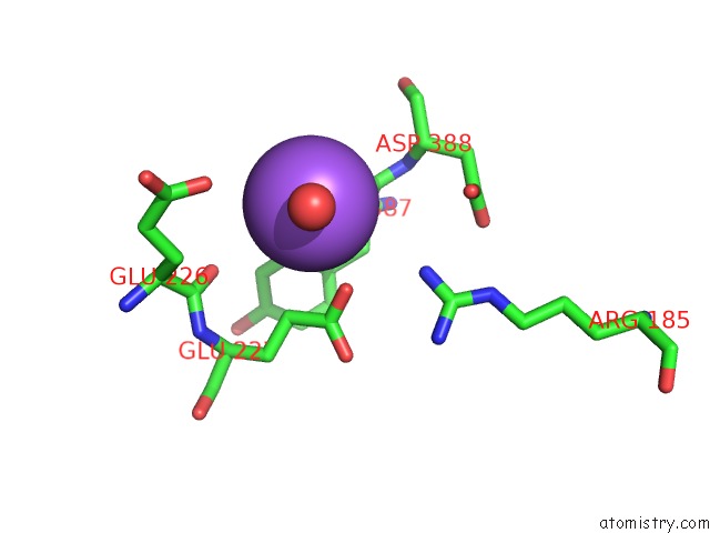
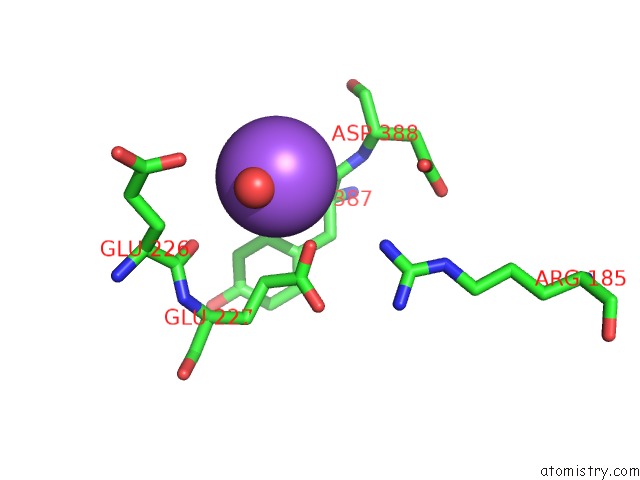
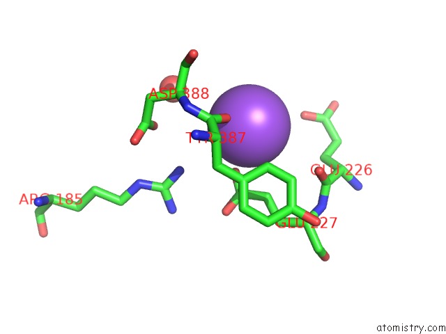
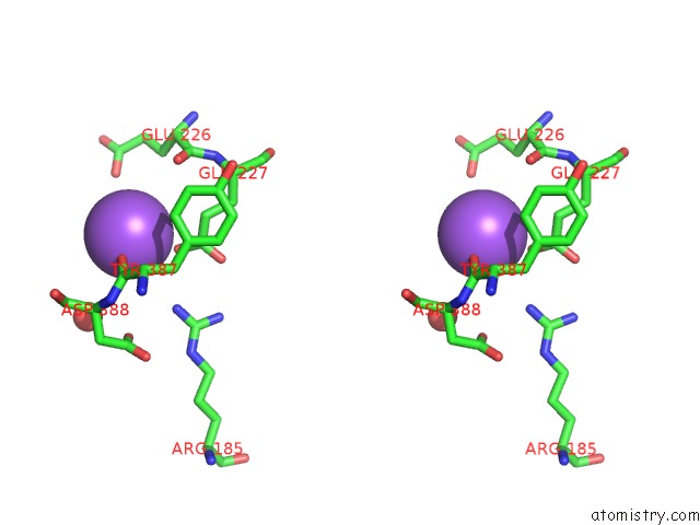
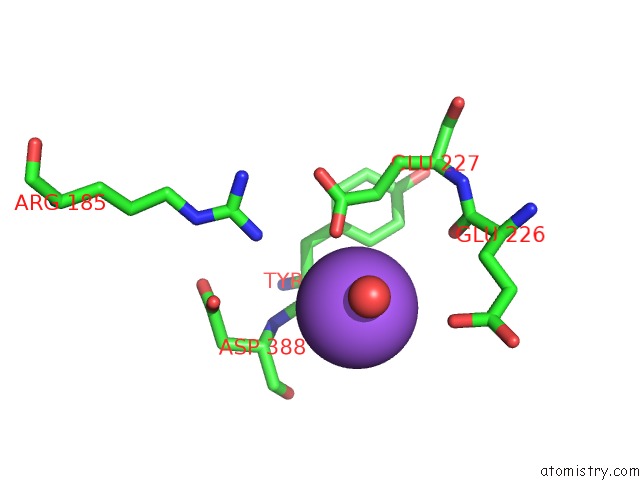
Sodium binding site 1 out of 6 in 3n7z
Go back to
Sodium binding site 1 out
of 6 in the Crystal Structure of Acetyltransferase From Bacillus Anthracis
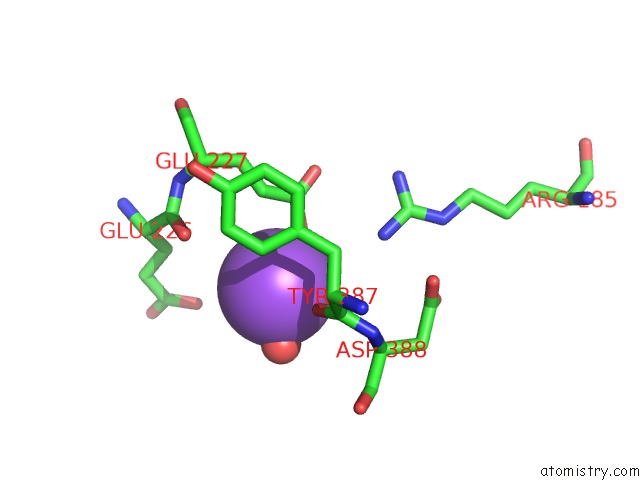
Mono view
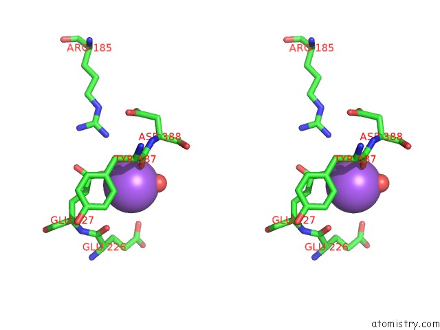
Stereo pair view
Mono view
Stereo pair view
A full contact list of Sodium with other atoms in the Na binding
site number 1 of Crystal Structure of Acetyltransferase From Bacillus Anthracis within 5.0Å range:
|
Sodium binding site 2 out of 6 in 3n7z
Go back to
Sodium binding site 2 out
of 6 in the Crystal Structure of Acetyltransferase From Bacillus Anthracis
Mono view
Stereo pair view
Mono view
Stereo pair view
A full contact list of Sodium with other atoms in the Na binding
site number 2 of Crystal Structure of Acetyltransferase From Bacillus Anthracis within 5.0Å range:
|
Sodium binding site 3 out of 6 in 3n7z
Go back to
Sodium binding site 3 out
of 6 in the Crystal Structure of Acetyltransferase From Bacillus Anthracis
Mono view

Stereo pair view
Mono view
Stereo pair view
A full contact list of Sodium with other atoms in the Na binding
site number 3 of Crystal Structure of Acetyltransferase From Bacillus Anthracis within 5.0Å range:
|
Sodium binding site 4 out of 6 in 3n7z
Go back to
Sodium binding site 4 out
of 6 in the Crystal Structure of Acetyltransferase From Bacillus Anthracis
Mono view
Stereo pair view
Mono view
Stereo pair view
A full contact list of Sodium with other atoms in the Na binding
site number 4 of Crystal Structure of Acetyltransferase From Bacillus Anthracis within 5.0Å range:
|
Sodium binding site 5 out of 6 in 3n7z
Go back to
Sodium binding site 5 out
of 6 in the Crystal Structure of Acetyltransferase From Bacillus Anthracis

Mono view
Stereo pair view
Mono view
Stereo pair view
A full contact list of Sodium with other atoms in the Na binding
site number 5 of Crystal Structure of Acetyltransferase From Bacillus Anthracis within 5.0Å range:
|
Sodium binding site 6 out of 6 in 3n7z
Go back to
Sodium binding site 6 out
of 6 in the Crystal Structure of Acetyltransferase From Bacillus Anthracis
Mono view
Stereo pair view
Mono view
Stereo pair view
A full contact list of Sodium with other atoms in the Na binding
site number 6 of Crystal Structure of Acetyltransferase From Bacillus Anthracis within 5.0Å range:
|
Reference:
K.D.Green,
T.Biswas,
C.Chang,
R.Wu,
W.Chen,
B.K.Janes,
D.Chalupska,
P.Gornicki,
P.C.Hanna,
O.V.Tsodikov,
A.Joachimiak,
S.Garneau-Tsodikova.
Biochemical and Structural Analysis of An Eis Family Aminoglycoside Acetyltransferase From Bacillus Anthracis. Biochemistry V. 54 3197 2015.
ISSN: ISSN 0006-2960
PubMed: 25928210
DOI: 10.1021/ACS.BIOCHEM.5B00244
Page generated: Mon Oct 7 11:44:43 2024
ISSN: ISSN 0006-2960
PubMed: 25928210
DOI: 10.1021/ACS.BIOCHEM.5B00244
Last articles
F in 7MB2F in 7MB1
F in 7MAX
F in 7M8R
F in 7M9R
F in 7M94
F in 7M8Q
F in 7M91
F in 7M8O
F in 7M8P