Sodium »
PDB 3moc-3n0p »
3mux »
Sodium in PDB 3mux: The Crystal Structure of A Putative 4-Hydroxy-2-Oxoglutarate Aldolase From Bacillus Anthracis to 1.45A
Protein crystallography data
The structure of The Crystal Structure of A Putative 4-Hydroxy-2-Oxoglutarate Aldolase From Bacillus Anthracis to 1.45A, PDB code: 3mux
was solved by
A.J.Stein,
C.Hatzos-Skintges,
S.Clancy,
A.Joachimiak,
Midwest Center Forstructural Genomics (Mcsg),
with X-Ray Crystallography technique. A brief refinement statistics is given in the table below:
| Resolution Low / High (Å) | 29.15 / 1.45 |
| Space group | P 21 21 21 |
| Cell size a, b, c (Å), α, β, γ (°) | 55.669, 68.431, 131.081, 90.00, 90.00, 90.00 |
| R / Rfree (%) | 14.8 / 17.9 |
Other elements in 3mux:
The structure of The Crystal Structure of A Putative 4-Hydroxy-2-Oxoglutarate Aldolase From Bacillus Anthracis to 1.45A also contains other interesting chemical elements:
| Chlorine | (Cl) | 3 atoms |
Sodium Binding Sites:
The binding sites of Sodium atom in the The Crystal Structure of A Putative 4-Hydroxy-2-Oxoglutarate Aldolase From Bacillus Anthracis to 1.45A
(pdb code 3mux). This binding sites where shown within
5.0 Angstroms radius around Sodium atom.
In total 2 binding sites of Sodium where determined in the The Crystal Structure of A Putative 4-Hydroxy-2-Oxoglutarate Aldolase From Bacillus Anthracis to 1.45A, PDB code: 3mux:
Jump to Sodium binding site number: 1; 2;
In total 2 binding sites of Sodium where determined in the The Crystal Structure of A Putative 4-Hydroxy-2-Oxoglutarate Aldolase From Bacillus Anthracis to 1.45A, PDB code: 3mux:
Jump to Sodium binding site number: 1; 2;
Sodium binding site 1 out of 2 in 3mux
Go back to
Sodium binding site 1 out
of 2 in the The Crystal Structure of A Putative 4-Hydroxy-2-Oxoglutarate Aldolase From Bacillus Anthracis to 1.45A
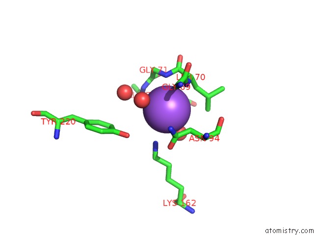
Mono view
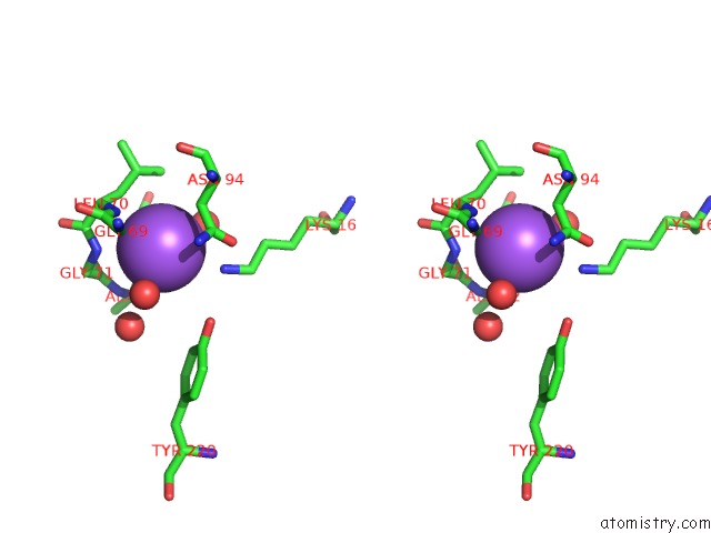
Stereo pair view
Mono view
Stereo pair view
A full contact list of Sodium with other atoms in the Na binding
site number 1 of The Crystal Structure of A Putative 4-Hydroxy-2-Oxoglutarate Aldolase From Bacillus Anthracis to 1.45A within 5.0Å range:
|
Sodium binding site 2 out of 2 in 3mux
Go back to
Sodium binding site 2 out
of 2 in the The Crystal Structure of A Putative 4-Hydroxy-2-Oxoglutarate Aldolase From Bacillus Anthracis to 1.45A
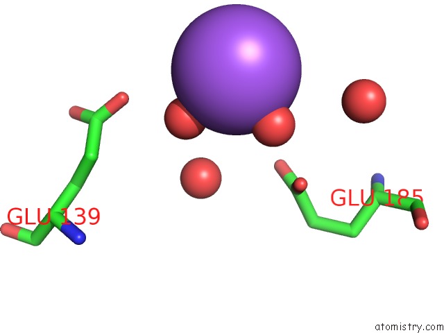
Mono view
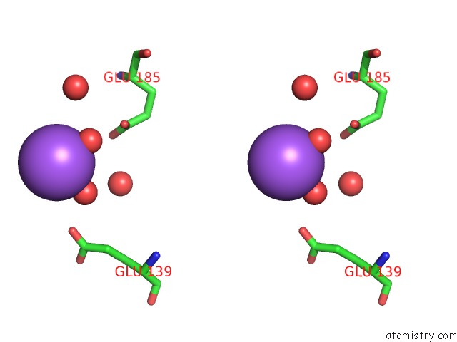
Stereo pair view
Mono view
Stereo pair view
A full contact list of Sodium with other atoms in the Na binding
site number 2 of The Crystal Structure of A Putative 4-Hydroxy-2-Oxoglutarate Aldolase From Bacillus Anthracis to 1.45A within 5.0Å range:
|
Reference:
A.J.Stein,
C.Hatzos-Skintges,
S.Clancy,
A.Joachimiak.
The Crystal Structure of A Putative 4-Hydroxy-2-Oxoglutarate Aldolase From Bacillus Anthracis to 1.45A To Be Published.
Page generated: Mon Oct 7 11:37:06 2024
Last articles
Cl in 7SX6Cl in 7STD
Cl in 7SWR
Cl in 7SVT
Cl in 7SUU
Cl in 7SUJ
Cl in 7SUT
Cl in 7SUI
Cl in 7SUH
Cl in 7SQE