Sodium »
PDB 3moc-3n0p »
3mr1 »
Sodium in PDB 3mr1: Crystal Structure of Methionine Aminopeptidase From Rickettsia Prowazekii
Enzymatic activity of Crystal Structure of Methionine Aminopeptidase From Rickettsia Prowazekii
All present enzymatic activity of Crystal Structure of Methionine Aminopeptidase From Rickettsia Prowazekii:
3.4.11.18;
3.4.11.18;
Protein crystallography data
The structure of Crystal Structure of Methionine Aminopeptidase From Rickettsia Prowazekii, PDB code: 3mr1
was solved by
Seattle Structural Genomics Center For Infectious Disease (Ssgcid),
with X-Ray Crystallography technique. A brief refinement statistics is given in the table below:
| Resolution Low / High (Å) | 42.40 / 2.00 |
| Space group | P 1 21 1 |
| Cell size a, b, c (Å), α, β, γ (°) | 42.480, 114.850, 115.800, 90.00, 92.66, 90.00 |
| R / Rfree (%) | 17.1 / 21.2 |
Other elements in 3mr1:
The structure of Crystal Structure of Methionine Aminopeptidase From Rickettsia Prowazekii also contains other interesting chemical elements:
| Manganese | (Mn) | 8 atoms |
| Chlorine | (Cl) | 2 atoms |
Sodium Binding Sites:
The binding sites of Sodium atom in the Crystal Structure of Methionine Aminopeptidase From Rickettsia Prowazekii
(pdb code 3mr1). This binding sites where shown within
5.0 Angstroms radius around Sodium atom.
In total 4 binding sites of Sodium where determined in the Crystal Structure of Methionine Aminopeptidase From Rickettsia Prowazekii, PDB code: 3mr1:
Jump to Sodium binding site number: 1; 2; 3; 4;
In total 4 binding sites of Sodium where determined in the Crystal Structure of Methionine Aminopeptidase From Rickettsia Prowazekii, PDB code: 3mr1:
Jump to Sodium binding site number: 1; 2; 3; 4;
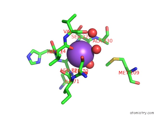
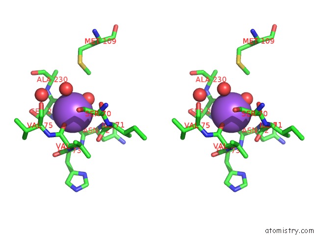
Sodium binding site 1 out of 4 in 3mr1
Go back to
Sodium binding site 1 out
of 4 in the Crystal Structure of Methionine Aminopeptidase From Rickettsia Prowazekii
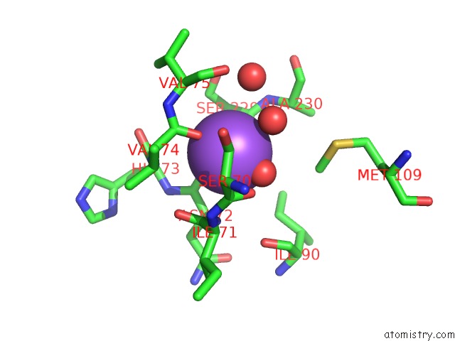
Mono view
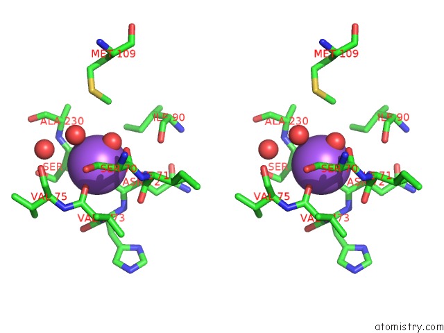
Stereo pair view
Mono view
Stereo pair view
A full contact list of Sodium with other atoms in the Na binding
site number 1 of Crystal Structure of Methionine Aminopeptidase From Rickettsia Prowazekii within 5.0Å range:
|
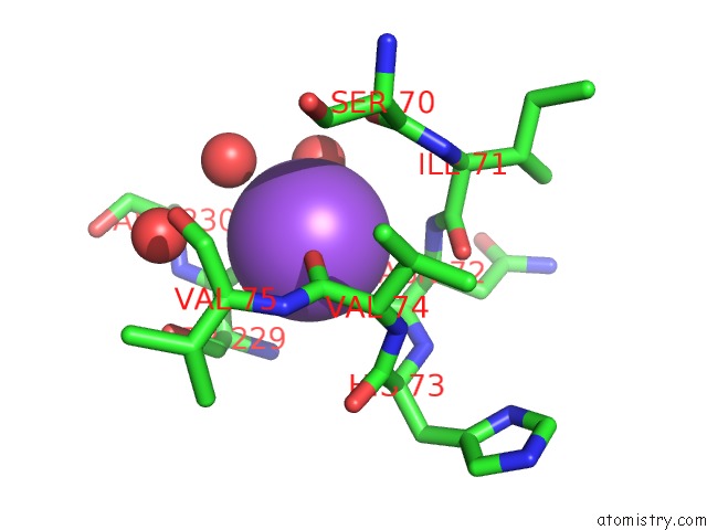
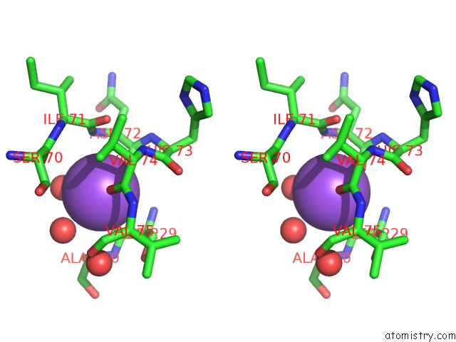
Sodium binding site 2 out of 4 in 3mr1
Go back to
Sodium binding site 2 out
of 4 in the Crystal Structure of Methionine Aminopeptidase From Rickettsia Prowazekii
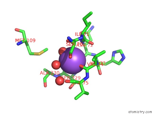
Mono view
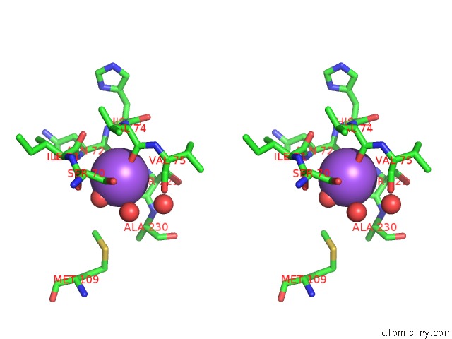
Stereo pair view
Mono view
Stereo pair view
A full contact list of Sodium with other atoms in the Na binding
site number 2 of Crystal Structure of Methionine Aminopeptidase From Rickettsia Prowazekii within 5.0Å range:
|
Sodium binding site 3 out of 4 in 3mr1
Go back to
Sodium binding site 3 out
of 4 in the Crystal Structure of Methionine Aminopeptidase From Rickettsia Prowazekii
Mono view
Stereo pair view
Mono view
Stereo pair view
A full contact list of Sodium with other atoms in the Na binding
site number 3 of Crystal Structure of Methionine Aminopeptidase From Rickettsia Prowazekii within 5.0Å range:
|
Sodium binding site 4 out of 4 in 3mr1
Go back to
Sodium binding site 4 out
of 4 in the Crystal Structure of Methionine Aminopeptidase From Rickettsia Prowazekii
Mono view
Stereo pair view
Mono view
Stereo pair view
A full contact list of Sodium with other atoms in the Na binding
site number 4 of Crystal Structure of Methionine Aminopeptidase From Rickettsia Prowazekii within 5.0Å range:
|
Reference:
T.R.Helgren,
C.Chen,
P.Wangtrakuldee,
T.E.Edwards,
B.L.Staker,
J.Abendroth,
B.Sankaran,
N.A.Housley,
P.J.Myler,
J.P.Audia,
J.R.Horn,
T.J.Hagen.
Rickettsia Prowazekii Methionine Aminopeptidase As A Promising Target For the Development of Antibacterial Agents. Bioorg.Med.Chem. V. 25 813 2017.
ISSN: ISSN 0968-0896
PubMed: 28089350
DOI: 10.1016/J.BMC.2016.11.013
Page generated: Mon Oct 7 11:35:33 2024
ISSN: ISSN 0968-0896
PubMed: 28089350
DOI: 10.1016/J.BMC.2016.11.013
Last articles
Zn in 9MJ5Zn in 9HNW
Zn in 9G0L
Zn in 9FNE
Zn in 9DZN
Zn in 9E0I
Zn in 9D32
Zn in 9DAK
Zn in 8ZXC
Zn in 8ZUF