Sodium »
PDB 3lbg-3m9y »
3m9y »
Sodium in PDB 3m9y: Crystal Structure of Triosephosphate Isomerase From Methicillin Resistant Staphylococcus Aureus at 1.9 Angstrom Resolution
Enzymatic activity of Crystal Structure of Triosephosphate Isomerase From Methicillin Resistant Staphylococcus Aureus at 1.9 Angstrom Resolution
All present enzymatic activity of Crystal Structure of Triosephosphate Isomerase From Methicillin Resistant Staphylococcus Aureus at 1.9 Angstrom Resolution:
5.3.1.1;
5.3.1.1;
Protein crystallography data
The structure of Crystal Structure of Triosephosphate Isomerase From Methicillin Resistant Staphylococcus Aureus at 1.9 Angstrom Resolution, PDB code: 3m9y
was solved by
S.Mukherjee,
D.Dutta,
B.Saha,
A.K.Das,
with X-Ray Crystallography technique. A brief refinement statistics is given in the table below:
| Resolution Low / High (Å) | 20.00 / 1.90 |
| Space group | P 43 21 2 |
| Cell size a, b, c (Å), α, β, γ (°) | 79.478, 79.478, 175.006, 90.00, 90.00, 90.00 |
| R / Rfree (%) | 15.9 / 20.5 |
Sodium Binding Sites:
The binding sites of Sodium atom in the Crystal Structure of Triosephosphate Isomerase From Methicillin Resistant Staphylococcus Aureus at 1.9 Angstrom Resolution
(pdb code 3m9y). This binding sites where shown within
5.0 Angstroms radius around Sodium atom.
In total 2 binding sites of Sodium where determined in the Crystal Structure of Triosephosphate Isomerase From Methicillin Resistant Staphylococcus Aureus at 1.9 Angstrom Resolution, PDB code: 3m9y:
Jump to Sodium binding site number: 1; 2;
In total 2 binding sites of Sodium where determined in the Crystal Structure of Triosephosphate Isomerase From Methicillin Resistant Staphylococcus Aureus at 1.9 Angstrom Resolution, PDB code: 3m9y:
Jump to Sodium binding site number: 1; 2;
Sodium binding site 1 out of 2 in 3m9y
Go back to
Sodium binding site 1 out
of 2 in the Crystal Structure of Triosephosphate Isomerase From Methicillin Resistant Staphylococcus Aureus at 1.9 Angstrom Resolution

Mono view
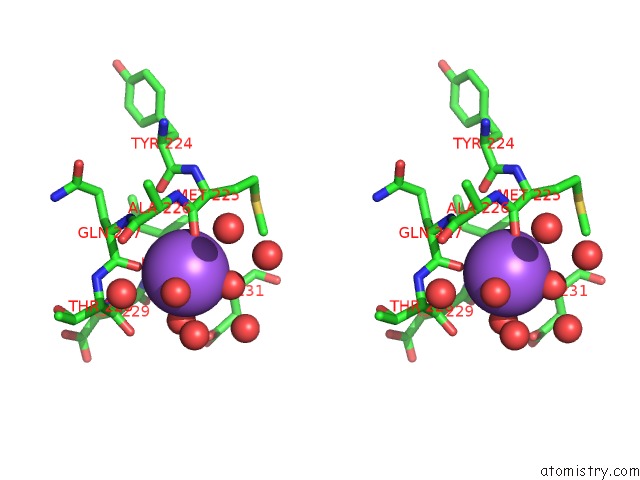
Stereo pair view
Mono view
Stereo pair view
A full contact list of Sodium with other atoms in the Na binding
site number 1 of Crystal Structure of Triosephosphate Isomerase From Methicillin Resistant Staphylococcus Aureus at 1.9 Angstrom Resolution within 5.0Å range:
|
Sodium binding site 2 out of 2 in 3m9y
Go back to
Sodium binding site 2 out
of 2 in the Crystal Structure of Triosephosphate Isomerase From Methicillin Resistant Staphylococcus Aureus at 1.9 Angstrom Resolution

Mono view

Stereo pair view
Mono view
Stereo pair view
A full contact list of Sodium with other atoms in the Na binding
site number 2 of Crystal Structure of Triosephosphate Isomerase From Methicillin Resistant Staphylococcus Aureus at 1.9 Angstrom Resolution within 5.0Å range:
|
Reference:
S.Mukherjee,
A.Roychowdhury,
D.Dutta,
A.K.Das.
Crystal Structures of Triosephosphate Isomerase From Methicillin Resistant Staphylococcus Aureus MRSA252 Provide Structural Insights Into Novel Modes of Ligand Binding and Unique Conformations of Catalytic Loop Biochimie V. 94 2532 2012.
ISSN: ISSN 0300-9084
PubMed: 22813930
DOI: 10.1016/J.BIOCHI.2012.07.001
Page generated: Mon Oct 7 11:26:28 2024
ISSN: ISSN 0300-9084
PubMed: 22813930
DOI: 10.1016/J.BIOCHI.2012.07.001
Last articles
Zn in 9MJ5Zn in 9HNW
Zn in 9G0L
Zn in 9FNE
Zn in 9DZN
Zn in 9E0I
Zn in 9D32
Zn in 9DAK
Zn in 8ZXC
Zn in 8ZUF