Sodium »
PDB 3fvs-3gdg »
3ga6 »
Sodium in PDB 3ga6: MTH0212 in Complex with Two Dna Helices
Enzymatic activity of MTH0212 in Complex with Two Dna Helices
All present enzymatic activity of MTH0212 in Complex with Two Dna Helices:
3.1.11.2;
3.1.11.2;
Protein crystallography data
The structure of MTH0212 in Complex with Two Dna Helices, PDB code: 3ga6
was solved by
K.Lakomek,
A.Dickmanns,
R.Ficner,
with X-Ray Crystallography technique. A brief refinement statistics is given in the table below:
| Resolution Low / High (Å) | 33.71 / 1.90 |
| Space group | P 1 21 1 |
| Cell size a, b, c (Å), α, β, γ (°) | 54.834, 126.655, 54.826, 90.00, 93.14, 90.00 |
| R / Rfree (%) | 15.5 / 20.3 |
Sodium Binding Sites:
The binding sites of Sodium atom in the MTH0212 in Complex with Two Dna Helices
(pdb code 3ga6). This binding sites where shown within
5.0 Angstroms radius around Sodium atom.
In total only one binding site of Sodium was determined in the MTH0212 in Complex with Two Dna Helices, PDB code: 3ga6:
In total only one binding site of Sodium was determined in the MTH0212 in Complex with Two Dna Helices, PDB code: 3ga6:
Sodium binding site 1 out of 1 in 3ga6
Go back to
Sodium binding site 1 out
of 1 in the MTH0212 in Complex with Two Dna Helices
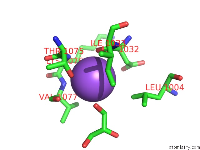
Mono view
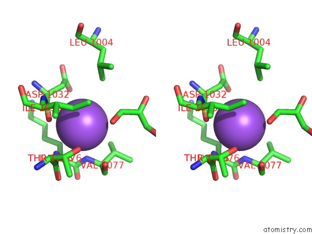
Stereo pair view
Mono view
Stereo pair view
A full contact list of Sodium with other atoms in the Na binding
site number 1 of MTH0212 in Complex with Two Dna Helices within 5.0Å range:
|
Reference:
K.Lakomek,
A.Dickmanns,
E.Ciirdaeva,
L.Schomacher,
R.Ficner.
Crystal Structure Analysis of Dna Uridine Endonuclease MTH212 Bound to Dna J.Mol.Biol. V. 399 604 2010.
ISSN: ISSN 0022-2836
PubMed: 20434457
DOI: 10.1016/J.JMB.2010.04.044
Page generated: Mon Oct 7 09:57:01 2024
ISSN: ISSN 0022-2836
PubMed: 20434457
DOI: 10.1016/J.JMB.2010.04.044
Last articles
Zn in 9MJ5Zn in 9HNW
Zn in 9G0L
Zn in 9FNE
Zn in 9DZN
Zn in 9E0I
Zn in 9D32
Zn in 9DAK
Zn in 8ZXC
Zn in 8ZUF