Sodium »
PDB 3fak-3fvi »
3fob »
Sodium in PDB 3fob: Crystal Structure of Bromoperoxidase From Bacillus Anthracis
Protein crystallography data
The structure of Crystal Structure of Bromoperoxidase From Bacillus Anthracis, PDB code: 3fob
was solved by
J.Osipiuk,
M.Gu,
J.Stam,
W.F.Anderson,
A.Joachimiak,
Center For Structuralgenomics Of Infectious Diseases (Csgid),
with X-Ray Crystallography technique. A brief refinement statistics is given in the table below:
| Resolution Low / High (Å) | 27.50 / 1.74 |
| Space group | P 1 21 1 |
| Cell size a, b, c (Å), α, β, γ (°) | 77.037, 48.326, 101.932, 90.00, 94.05, 90.00 |
| R / Rfree (%) | 16.4 / 19.5 |
Other elements in 3fob:
The structure of Crystal Structure of Bromoperoxidase From Bacillus Anthracis also contains other interesting chemical elements:
| Chlorine | (Cl) | 3 atoms |
Sodium Binding Sites:
The binding sites of Sodium atom in the Crystal Structure of Bromoperoxidase From Bacillus Anthracis
(pdb code 3fob). This binding sites where shown within
5.0 Angstroms radius around Sodium atom.
In total 2 binding sites of Sodium where determined in the Crystal Structure of Bromoperoxidase From Bacillus Anthracis, PDB code: 3fob:
Jump to Sodium binding site number: 1; 2;
In total 2 binding sites of Sodium where determined in the Crystal Structure of Bromoperoxidase From Bacillus Anthracis, PDB code: 3fob:
Jump to Sodium binding site number: 1; 2;
Sodium binding site 1 out of 2 in 3fob
Go back to
Sodium binding site 1 out
of 2 in the Crystal Structure of Bromoperoxidase From Bacillus Anthracis
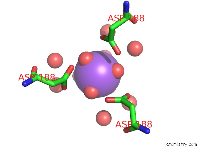
Mono view
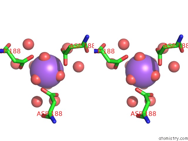
Stereo pair view
Mono view
Stereo pair view
A full contact list of Sodium with other atoms in the Na binding
site number 1 of Crystal Structure of Bromoperoxidase From Bacillus Anthracis within 5.0Å range:
|
Sodium binding site 2 out of 2 in 3fob
Go back to
Sodium binding site 2 out
of 2 in the Crystal Structure of Bromoperoxidase From Bacillus Anthracis
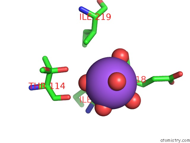
Mono view
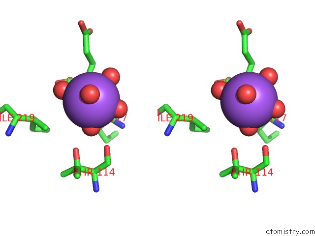
Stereo pair view
Mono view
Stereo pair view
A full contact list of Sodium with other atoms in the Na binding
site number 2 of Crystal Structure of Bromoperoxidase From Bacillus Anthracis within 5.0Å range:
|
Reference:
J.Osipiuk,
M.Gu,
J.Stam,
W.F.Anderson,
A.Joachimiak.
X-Ray Crystal Structure of Bromoperoxidase From Bacillus Anthracis. To Be Published.
Page generated: Sun Aug 17 14:51:11 2025
Last articles
Na in 5EEMNa in 5DYN
Na in 5EDF
Na in 5EB8
Na in 5EBA
Na in 5E9R
Na in 5E8B
Na in 5E9N
Na in 5E7H
Na in 5E8E